• 文献检索
  • 文档翻译
  • 深度研究
  • 学术资讯
  • Suppr Zotero 插件Zotero 插件
  • 邀请有礼
  • 套餐&价格
  • 历史记录
应用&插件
Suppr Zotero 插件Zotero 插件浏览器插件Mac 客户端Windows 客户端微信小程序
定价
高级版会员购买积分包购买API积分包
服务
文献检索文档翻译深度研究API 文档MCP 服务
关于我们
关于 Suppr公司介绍联系我们用户协议隐私条款
关注我们

Suppr 超能文献

核心技术专利:CN118964589B侵权必究
粤ICP备2023148730 号-1Suppr @ 2026

文献检索

告别复杂PubMed语法,用中文像聊天一样搜索,搜遍4000万医学文献。AI智能推荐,让科研检索更轻松。

立即免费搜索

文件翻译

保留排版,准确专业,支持PDF/Word/PPT等文件格式,支持 12+语言互译。

免费翻译文档

深度研究

AI帮你快速写综述,25分钟生成高质量综述,智能提取关键信息,辅助科研写作。

立即免费体验

非小细胞肺癌患者的脑成像——一项系统综述

Brain Imaging in Patients with Non-Small Cell Lung Cancer-A Systematic Review.

作者信息

Mayer Nora, Boschetti Laura, Scarci Marco, Cioffi Ugo, De Simone Matilde, Schnider Marlène, Kestenholz Peter, Minervini Fabrizio

机构信息

Division of Thoracic Surgery, Cantonal Hospital Lucerne, 6000 Lucerne, Switzerland.

Department of Medical Oncology, Cantonal Hospital Lucerne, 6210 Sursee, Switzerland.

出版信息

J Clin Med. 2025 Jan 22;14(3):708. doi: 10.3390/jcm14030708.

DOI:10.3390/jcm14030708
PMID:39941379
原文链接:https://pmc.ncbi.nlm.nih.gov/articles/PMC11818832/
Abstract

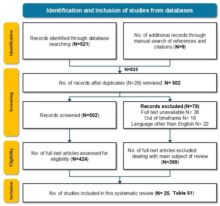
: Lung cancer frequently metastasizes to the brain, liver, and adrenal glands with a significant negative prognostic impact on overall survival and quality of life (QoL). To optimize treatment and prognosis, adequate staging with the detection of distant metastases is crucial. The incidence of brain metastases in potentially resectable early-stage non-small cell lung cancer (NSCLC) is as low as 3%; hence, the need for preoperative brain imaging has been a constant matter of debate, especially in stage II. In stages III and IV NSCLC, neuroimaging is an essential part of staging. : A systematic literature search was performed. Publications from 1999 to 2024, focusing on preoperative brain imaging (BI) in the staging of stages I-IV NSCLC, were included. Data extraction included study population characteristics, the modality of BI, the incidence of brain metastases (BMs), and the main outcomes of the studies. The final included studies were selected according to the PRISMA criteria. In the second step, guidelines on BI in NSCLC staging of major importance were identified and compared. : A total of 530 articles were identified, of which 25 articles were selected. Four prospective studies and 21 retrospective investigations were included. Most of the investigations focused on BI in the early stages. The main imaging modality for BI was magnetic resonance imaging (MRI), followed by computed tomography (CT). Besides the identified 25 studies, the most important internationally applied guidelines on brain imaging in the staging of NSCLC were reviewed. While some guidelines agree on preoperative BI in NSCLC stage III (Union for International Cancer Control-UICC eighth edition) patients, other guidelines recommend earlier BI starting from clinical stage II. All mentioned guidelines homogenously recommend BI in patients with symptoms suggestive of brain pathologies. : BI in NSCLC staging is recommended in neurologically symptomatic patients suggestive of brain metastases as well as NSCLC patients with stage III disease. Neuroimaging in stage IA patients, as well as in pure GGO (Ground-Glass Opacity) lesions, was considered unnecessary. The predominantly applied imaging modality was ce-MRI (contrast-enhanced magnetic resonance imaging). Inconsistency exists concerning BI in stage II. The identification of prognostic factors for developing BM in patients with early-stage NSCLC could help to clarify which subgroup might benefit from preoperative BI.

摘要

肺癌常转移至脑、肝和肾上腺,对总生存期和生活质量(QoL)有显著的负面预后影响。为优化治疗和预后,通过检测远处转移进行充分分期至关重要。在潜在可切除的早期非小细胞肺癌(NSCLC)中,脑转移的发生率低至3%;因此,术前脑成像的必要性一直是争论的焦点,尤其是在II期。在III期和IV期NSCLC中,神经成像检查是分期的重要组成部分。

进行了系统的文献检索。纳入了1999年至2024年期间聚焦于I-IV期NSCLC分期中术前脑成像(BI)的出版物。数据提取包括研究人群特征、BI的方式、脑转移(BMs)的发生率以及研究的主要结果。最终纳入的研究根据PRISMA标准进行选择。第二步,确定并比较了NSCLC分期中关于BI的重要指南。

共识别出530篇文章,其中25篇被选中。包括4项前瞻性研究和21项回顾性调查。大多数研究聚焦于早期的BI。BI的主要成像方式是磁共振成像(MRI),其次是计算机断层扫描(CT)。除了已识别的25项研究外,还回顾了NSCLC分期中最重要的国际应用脑成像指南。虽然一些指南对于NSCLC III期(国际癌症控制联盟-UICC第八版)患者的术前BI意见一致,但其他指南建议从临床II期开始更早进行BI。所有提及的指南均一致推荐对有脑病变症状的患者进行BI。

对于有脑转移提示症状的神经症状患者以及III期疾病的NSCLC患者,建议在NSCLC分期中进行BI。对于IA期患者以及纯磨玻璃密度(GGO)病变患者,认为无需进行神经成像检查。主要应用的成像方式是增强磁共振成像(ce-MRI)。关于II期的BI存在不一致之处。识别早期NSCLC患者发生BM的预后因素有助于明确哪些亚组可能从术前BI中获益。

https://cdn.ncbi.nlm.nih.gov/pmc/blobs/a940/11818832/bf8ef2d0f58a/jcm-14-00708-g004.jpg
https://cdn.ncbi.nlm.nih.gov/pmc/blobs/a940/11818832/aed943a9787c/jcm-14-00708-g001.jpg
https://cdn.ncbi.nlm.nih.gov/pmc/blobs/a940/11818832/a24cb7aafb72/jcm-14-00708-g002.jpg
https://cdn.ncbi.nlm.nih.gov/pmc/blobs/a940/11818832/e8d4cad3527c/jcm-14-00708-g003.jpg
https://cdn.ncbi.nlm.nih.gov/pmc/blobs/a940/11818832/bf8ef2d0f58a/jcm-14-00708-g004.jpg

相似文献

1
Brain Imaging in Patients with Non-Small Cell Lung Cancer-A Systematic Review.非小细胞肺癌患者的脑成像——一项系统综述
J Clin Med. 2025 Jan 22;14(3):708. doi: 10.3390/jcm14030708.
2
Screening for brain metastases in patients with stage III non-small-cell lung cancer, magnetic resonance imaging or computed tomography? A prospective study.III 期非小细胞肺癌患者脑转移的筛查:磁共振成像还是计算机断层扫描?一项前瞻性研究。
Eur J Cancer. 2019 Jul;115:88-96. doi: 10.1016/j.ejca.2019.04.017. Epub 2019 May 23.
3
Screening for brain metastases in patients with stage III non-small cell lung cancer: Is there additive value of magnetic resonance imaging above a contrast-enhanced computed tomography of the brain?III 期非小细胞肺癌患者脑转移筛查:磁共振成像相对于增强 CT 扫描是否具有附加价值?
Lung Cancer. 2013 Jun;80(3):293-7. doi: 10.1016/j.lungcan.2013.02.006. Epub 2013 Mar 18.
4
Significance of Brain Imaging for Staging in Patients With Clinical Stage T1-2 N0 Non-Small-Cell Lung Cancer on Positron Emission Tomography/Computed Tomography.正电子发射断层扫描/计算机断层扫描临床 T1-2N0 期非小细胞肺癌患者脑影像学分期的意义。
Clin Lung Cancer. 2021 Nov;22(6):562-569. doi: 10.1016/j.cllc.2021.06.004. Epub 2021 Jun 13.
5
Routine preoperative brain CT in resectable non-small cell lung cancer - Ten years experience from a tertiary UK thoracic center.可切除非小细胞肺癌患者的常规术前脑部 CT-英国一家三级胸科中心的十年经验。
Lung Cancer. 2018 Aug;122:195-199. doi: 10.1016/j.lungcan.2018.06.014. Epub 2018 Jun 10.
6
Non-small cell lung cancer brain metastasis screening in the era of positron emission tomography-CT staging: Current practice and outcomes.正电子发射断层扫描-CT分期时代的非小细胞肺癌脑转移筛查:当前实践与结果
J Med Imaging Radiat Oncol. 2018 Jun;62(3):383-388. doi: 10.1111/1754-9485.12732. Epub 2018 May 10.
7
More advantages in detecting bone and soft tissue metastases from prostate cancer using F-PSMA PET/CT.使用F-PSMA PET/CT检测前列腺癌骨和软组织转移方面有更多优势。
Hell J Nucl Med. 2019 Jan-Apr;22(1):6-9. doi: 10.1967/s002449910952. Epub 2019 Mar 7.
8
Ultrasound, CT, MRI, or PET-CT for staging and re-staging of adults with cutaneous melanoma.超声、CT、MRI或PET-CT用于成人皮肤黑色素瘤的分期及再分期。
Cochrane Database Syst Rev. 2019 Jul 1;7(7):CD012806. doi: 10.1002/14651858.CD012806.pub2.
9
Consistency and prognostic value of preoperative staging and postoperative pathological staging using F-FDG PET/MRI in patients with non-small cell lung cancer.正电子发射断层扫描/磁共振成像(F-FDG PET/MRI)在非小细胞肺癌患者术前分期和术后病理分期中的一致性和预后价值。
Ann Nucl Med. 2022 Dec;36(12):1059-1072. doi: 10.1007/s12149-022-01795-9. Epub 2022 Oct 20.
10
Lymph Node Size Predicts for Asymptomatic Brain Metastases in Patients With Non-small-cell Lung Cancer at Diagnosis.淋巴结大小可预测非小细胞肺癌患者在诊断时的无症状脑转移。
Clin Lung Cancer. 2019 Jan;20(1):e107-e114. doi: 10.1016/j.cllc.2018.09.014. Epub 2018 Sep 24.

引用本文的文献

1
Machine learning-based radiomics for differentiating lung cancer subtypes in brain metastases using CE-T1WI.基于机器学习的影像组学在使用对比增强T1加权成像鉴别脑转移瘤中肺癌亚型的应用
Front Oncol. 2025 Jun 19;15:1599882. doi: 10.3389/fonc.2025.1599882. eCollection 2025.

本文引用的文献

1
Brain imaging in lung cancer staging: A real-world, multi-centre study of prevalence of brain metastases, impact on treatment and re-modelling of the NICE health economic analysis.肺癌分期中的脑成像:一项关于脑转移瘤患病率、对治疗的影响以及对英国国家卫生与临床优化研究所(NICE)健康经济分析重新建模的真实世界、多中心研究。
Lung Cancer. 2023 Apr;178:145-150. doi: 10.1016/j.lungcan.2023.02.012. Epub 2023 Feb 18.
2
Role of Pre-Operative Brain Imaging in Patients with NSCLC Stage I: A Retrospective, Multicenter Analysis.术前脑成像在非小细胞肺癌I期患者中的作用:一项回顾性多中心分析
Cancers (Basel). 2022 May 13;14(10):2419. doi: 10.3390/cancers14102419.
3
Revisiting Indications for Brain Imaging During the Clinical Staging Evaluation of Lung Cancer.
重新审视肺癌临床分期评估期间脑成像的指征
JTO Clin Res Rep. 2022 Apr 6;3(5):100318. doi: 10.1016/j.jtocrr.2022.100318. eCollection 2022 May.
4
No Prognostic Impact of Staging Brain MRI in Patients with Stage IA Non-Small Cell Lung Cancer.ⅠA 期非小细胞肺癌患者的分期脑 MRI 检查对预后无影响。
Radiology. 2022 Jun;303(3):632-643. doi: 10.1148/radiol.212101. Epub 2022 Mar 8.
5
Adjuvant atezolizumab after adjuvant chemotherapy in resected stage IB-IIIA non-small-cell lung cancer (IMpower010): a randomised, multicentre, open-label, phase 3 trial.辅助阿特珠单抗治疗辅助化疗后切除的 IB-IIIA 期非小细胞肺癌(IMpower010):一项随机、多中心、开放标签、III 期临床试验。
Lancet. 2021 Oct 9;398(10308):1344-1357. doi: 10.1016/S0140-6736(21)02098-5. Epub 2021 Sep 20.
6
Significance of Brain Imaging for Staging in Patients With Clinical Stage T1-2 N0 Non-Small-Cell Lung Cancer on Positron Emission Tomography/Computed Tomography.正电子发射断层扫描/计算机断层扫描临床 T1-2N0 期非小细胞肺癌患者脑影像学分期的意义。
Clin Lung Cancer. 2021 Nov;22(6):562-569. doi: 10.1016/j.cllc.2021.06.004. Epub 2021 Jun 13.
7
Development of Brain Metastases in Patients With Non-Small Cell Lung Cancer and No Brain Metastases at Initial Staging Evaluation: Cumulative Incidence and Risk Factor Analysis.初诊时无脑转移的非小细胞肺癌患者发生脑转移的发展:累积发生率和风险因素分析。
AJR Am J Roentgenol. 2021 Nov;217(5):1184-1193. doi: 10.2214/AJR.21.25787. Epub 2021 May 26.
8
Assessment of Brain Metastasis at Diagnosis in Non-Small-Cell Lung Cancer: A Prospective Observational Study From North India.诊断时非小细胞肺癌脑转移的评估:来自印度北部的一项前瞻性观察研究。
JCO Glob Oncol. 2021 Apr;7:593-601. doi: 10.1200/GO.20.00629.
9
The PRISMA 2020 statement: an updated guideline for reporting systematic reviews.PRISMA 2020 声明:系统评价报告的更新指南。
BMJ. 2021 Mar 29;372:n71. doi: 10.1136/bmj.n71.
10
Diagnostic Yield of Staging Brain MRI in Patients with Newly Diagnosed Non-Small Cell Lung Cancer.诊断性分期脑 MRI 在新诊断非小细胞肺癌患者中的应用。
Radiology. 2020 Nov;297(2):419-427. doi: 10.1148/radiol.2020201194. Epub 2020 Aug 25.