• 文献检索
  • 文档翻译
  • 深度研究
  • 学术资讯
  • Suppr Zotero 插件Zotero 插件
  • 邀请有礼
  • 套餐&价格
  • 历史记录
应用&插件
Suppr Zotero 插件Zotero 插件浏览器插件Mac 客户端Windows 客户端微信小程序
定价
高级版会员购买积分包购买API积分包
服务
文献检索文档翻译深度研究API 文档MCP 服务
关于我们
关于 Suppr公司介绍联系我们用户协议隐私条款
关注我们

Suppr 超能文献

核心技术专利:CN118964589B侵权必究
粤ICP备2023148730 号-1Suppr @ 2026

文献检索

告别复杂PubMed语法,用中文像聊天一样搜索,搜遍4000万医学文献。AI智能推荐,让科研检索更轻松。

立即免费搜索

文件翻译

保留排版,准确专业,支持PDF/Word/PPT等文件格式,支持 12+语言互译。

免费翻译文档

深度研究

AI帮你快速写综述,25分钟生成高质量综述,智能提取关键信息,辅助科研写作。

立即免费体验

优化多受试者内耳磁共振成像的空间归一化:不同几何保留配准方法的比较

Optimizing spatial normalization of multisubject inner ear MRI: comparison of different geometry-preserving co-registration approaches.

作者信息

Gerb Johannes, Kirsch Valerie, Kierig Emilie, Brandt Thomas, Dieterich Marianne, Boegle Rainer

机构信息

German Center for Vertigo and Balance Disorders (DSGZ), LMU University Hospital, Ludwig-Maximilians-University (LMU) Munich, Marchioninistraße 15, 81377, Munich, Germany.

Department of Neurology, LMU University Hospital, LMU Munich, Munich, Germany.

出版信息

Sci Rep. 2025 Feb 21;15(1):6414. doi: 10.1038/s41598-025-90842-2.

DOI:10.1038/s41598-025-90842-2
PMID:39984604
原文链接:https://pmc.ncbi.nlm.nih.gov/articles/PMC11845522/
Abstract

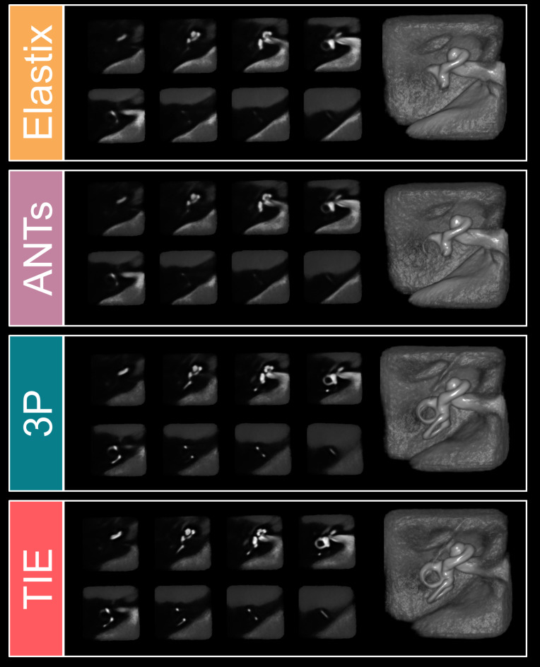
Spatial normalization of multisubject inner ear imaging data is challenging, due to both substantial intraindividual differences and the small size of the organ compared to other intracranial structures. Automatic whole brain co-registration to standard space can only roughly co-align the peripheral vestibular endorgan, and complemental manual registration is highly time-consuming. Here, we compared the accuracy of four geometry-maintaining co-registration methods (one semi-manual method and three automatic methods). High-resolution structural T2-MRI of 153 inner ears from patients and healthy participants were co-registered to an inner-ear atlas. The semi-manual method used a three-point landmark-based approach (3P), two automatic methods were based on unassisted standard algorithms (Advanced Normalization Tools (ANTs), Elastix (EL)), while the fourth automatic method utilized a volumetrically dilated, atlas-based mask (thick inner ear, TIE) for probabilistic inner ear masking. Registration accuracy was evaluated by neurotologists blinded to the respective registration paradigm, and the resulting median volumes were quantified using colocalization analyses. The mask-aided automatic approach showed the best ratings, followed by the semi-manual three-point landmark-based registration (mean ratings (lower: better) TIE 2.21 ± 1.15; 3P 2.58 ± 0.61; EL 3.42 ± 1.06; ANTs 3.49 ± 1.26). The semi-manual method had the lowest rate of insufficient registrations, followed by TIE (3P: 3.70%; TIE: 8.28%; EL: 22.66%; ANTs: 27.02%). TIE showed the highest colocalization metrics with the atlas. Only TIE and 3P allowed for sufficient semicircular canal visualization in method-wise average volumes. Overall, geometry-preserving spatial normalization of multisubject inner ear imaging data is possible and could allow groupwise examinations of the bony labyrinth or temporal bone morphology in the future.

摘要

多主体内耳成像数据的空间归一化具有挑战性,这是由于个体内部存在显著差异,且与其他颅内结构相比,该器官体积较小。自动全脑与标准空间的配准只能大致对齐外周前庭终器,而补充的手动配准非常耗时。在此,我们比较了四种保持几何形状的配准方法(一种半自动方法和三种自动方法)的准确性。对来自患者和健康参与者的153个内耳的高分辨率结构T2-MRI进行了与内耳图谱的配准。半自动方法采用基于三点地标法(3P),两种自动方法基于无辅助的标准算法(高级归一化工具(ANTs)、弹性配准软件(EL)),而第四种自动方法利用基于图谱的体积扩张掩码(厚内耳,TIE)进行概率性内耳掩膜。由对各自配准范式不知情的神经耳科医生评估配准准确性,并使用共定位分析对所得的中位数体积进行量化。掩码辅助自动方法的评分最佳,其次是基于三点地标的半自动配准(平均评分(越低越好):TIE 2.21±1.15;3P 2.58±0.61;EL 3.42±1.06;ANTs 3.49±1.26)。半自动方法的配准不足率最低,其次是TIE(3P:3.70%;TIE:8.28%;EL:22.66%;ANTs:27.02%)。TIE与图谱的共定位指标最高。仅TIE和3P在方法平均体积中能实现足够的半规管可视化。总体而言,多主体内耳成像数据的保持几何形状的空间归一化是可行的,并且未来可能允许对骨迷路或颞骨形态进行分组检查。

https://cdn.ncbi.nlm.nih.gov/pmc/blobs/9cdd/11845522/76c70f6cec85/41598_2025_90842_Fig5_HTML.jpg
https://cdn.ncbi.nlm.nih.gov/pmc/blobs/9cdd/11845522/f9cf11fc360a/41598_2025_90842_Fig1_HTML.jpg
https://cdn.ncbi.nlm.nih.gov/pmc/blobs/9cdd/11845522/52887f5da104/41598_2025_90842_Fig2_HTML.jpg
https://cdn.ncbi.nlm.nih.gov/pmc/blobs/9cdd/11845522/f4fcf545e407/41598_2025_90842_Fig3_HTML.jpg
https://cdn.ncbi.nlm.nih.gov/pmc/blobs/9cdd/11845522/bcb42f1c30a9/41598_2025_90842_Fig4_HTML.jpg
https://cdn.ncbi.nlm.nih.gov/pmc/blobs/9cdd/11845522/76c70f6cec85/41598_2025_90842_Fig5_HTML.jpg

相似文献

1
Optimizing spatial normalization of multisubject inner ear MRI: comparison of different geometry-preserving co-registration approaches.优化多受试者内耳磁共振成像的空间归一化:不同几何保留配准方法的比较
Sci Rep. 2025 Feb 21;15(1):6414. doi: 10.1038/s41598-025-90842-2.
2
A probabilistic atlas of the human inner ear's bony labyrinth enables reliable atlas-based segmentation of the total fluid space.人体内耳骨迷路概率图谱可实现总液腔基于图谱的可靠分割。
J Neurol. 2019 Sep;266(Suppl 1):52-61. doi: 10.1007/s00415-019-09488-6. Epub 2019 Aug 17.
3
Validation of inner ear MRI in patients with Ménière's disease by comparing endolymphatic hydrops from histopathologic specimens.通过比较组织病理学标本中的内淋巴积水来验证梅尼埃病患者的内耳 MRI。
Sci Rep. 2021 Sep 6;11(1):17738. doi: 10.1038/s41598-021-97213-7.
4
Automatic measurement of the labyrinth using image registration and a deformable inner ear atlas.使用图像配准和可变形内耳图谱自动测量迷路。
Acad Radiol. 2003 Sep;10(9):988-99. doi: 10.1016/s1076-6332(03)00121-1.
5
Results in caloric test, video head impulse test and inner ear MRI in patients with Ménière's disease.梅尼埃病患者的冷热试验、视频头脉冲试验及内耳磁共振成像结果。
Auris Nasus Larynx. 2020 Feb;47(1):71-78. doi: 10.1016/j.anl.2019.06.002. Epub 2019 Jul 2.
6
VOLT: a novel open-source pipeline for automatic segmentation of endolymphatic space in inner ear MRI.VOLT:一种新型的开源内耳膜迷路 MRI 内淋巴管自动分割流水线。
J Neurol. 2020 Dec;267(Suppl 1):185-196. doi: 10.1007/s00415-020-10062-8. Epub 2020 Jul 14.
7
Magnetic resonance imaging of endolymphatic hydrops in patients with unilateral Meniere's disease: a comparison between with and without herniation into the posterior and lateral semi-circular canals.单侧梅尼埃病患者内淋巴积水的磁共振成像:有无疝入后、外半规管的比较。
Acta Otolaryngol. 2021 Jul;141(7):671-677. doi: 10.1080/00016489.2021.1928282. Epub 2021 Jun 1.
8
The vestibular aqueduct ossification on temporal bone CT: an old sign revisited to rule out the presence of endolymphatic hydrops in Menière's disease patients.颞骨 CT 显示前庭水管骨化:重新审视这一古老征象,以排除梅尼埃病患者存在内淋巴积水。
Eur Radiol. 2020 Nov;30(11):6331-6338. doi: 10.1007/s00330-020-06980-w. Epub 2020 Jun 15.
9
[Application of 3D-Flair MRI and vestibular function assessment in profound sudden sensorineural hearing loss patients].3D-Flair磁共振成像及前庭功能评估在极重度突发性感音神经性听力损失患者中的应用
Zhonghua Er Bi Yan Hou Tou Jing Wai Ke Za Zhi. 2025 Jan 7;60(1):2-9. doi: 10.3760/cma.j.cn115330-20240403-00199.
10
Volume measurements of individual muscles in human quadriceps femoris using atlas-based segmentation approaches.使用基于图谱的分割方法对人股四头肌中的单个肌肉进行体积测量。
MAGMA. 2016 Apr;29(2):245-57. doi: 10.1007/s10334-016-0535-6. Epub 2016 Mar 16.

引用本文的文献

1
Contrast Agent Uptake in Endolymphatic Sac and Duct: Inverse Relation to Endolymphatic Hydrops.造影剂在内淋巴囊和内淋巴管中的摄取:与内淋巴积水呈负相关。
Laryngoscope. 2025 Aug;135(8):2914-2922. doi: 10.1002/lary.32127. Epub 2025 Mar 19.

本文引用的文献

1
The impact of 3D real-IR delayed post gadolinium MRI parameterisation on the diagnostic performance and optimal descriptor selection in Ménière's disease.三维真实红外延迟钆增强磁共振成像参数化对梅尼埃病诊断性能及最佳描述符选择的影响
Eur Radiol. 2025 Mar;35(3):1290-1302. doi: 10.1007/s00330-024-11218-0. Epub 2024 Dec 3.
2
Definition of a coordinate system for multi-modal images of the temporal bone and inner ear.颞骨和内耳多模态图像的坐标系定义。
PLoS One. 2024 Oct 7;19(10):e0294828. doi: 10.1371/journal.pone.0294828. eCollection 2024.
3
Imaging endolymphatic space of the inner ear in vestibular migraine.
前庭性偏头痛内耳内淋巴间隙的影像学表现
J Neurol Neurosurg Psychiatry. 2025 Apr 10;96(5):415-423. doi: 10.1136/jnnp-2024-334419.
4
Assessing the optimal MRI descriptors to diagnose Ménière's disease and the added value of analysing the vestibular aqueduct.评估诊断梅尼埃病的最佳 MRI 描述符及分析前庭水管的附加价值。
Eur Radiol. 2024 Sep;34(9):6060-6071. doi: 10.1007/s00330-024-10587-w. Epub 2024 Feb 7.
5
Towards fully automated inner ear analysis with deep-learning-based joint segmentation and landmark detection framework.基于深度学习的联合分割和地标检测框架实现内耳的全自动分析。
Sci Rep. 2023 Nov 4;13(1):19057. doi: 10.1038/s41598-023-45466-9.
6
Differential effects of gender and age on dynamic subjective visual vertical.性别和年龄对动态主观视觉垂直的差异影响。
Neurol Res Pract. 2023 Aug 24;5(1):37. doi: 10.1186/s42466-023-00266-4.
7
The spectrum of cochlear malformations in CHARGE syndrome and insights into the role of the CHD7 gene during embryogenesis of the inner ear.CHARGE 综合征中的耳蜗畸形谱及 CHD7 基因在内耳胚胎发育中的作用。
Neuroradiology. 2023 Apr;65(4):819-834. doi: 10.1007/s00234-023-03118-9. Epub 2023 Jan 30.
8
IE-Vnet: Deep Learning-Based Segmentation of the Inner Ear's Total Fluid Space.IE-Vnet:基于深度学习的内耳全液腔分割
Front Neurol. 2022 May 11;13:663200. doi: 10.3389/fneur.2022.663200. eCollection 2022.
9
Non-contrast MRI of Inner Ear Detected Differences of Endolymphatic Drainage System Between Vestibular Migraine and Unilateral Ménière's Disease.内耳非增强磁共振成像检测前庭性偏头痛与单侧梅尼埃病内淋巴引流系统的差异
Front Neurol. 2022 Apr 29;13:814518. doi: 10.3389/fneur.2022.814518. eCollection 2022.
10
Automatic intra-subject registration and fusion of multimodal cochlea 3D clinical images.多模态耳蜗三维临床图像的自动受试者内配准与融合。
PLoS One. 2022 Mar 2;17(3):e0264449. doi: 10.1371/journal.pone.0264449. eCollection 2022.