Hwang Sein, Sung Se In, Kim Young Eun, Yang Misun, Koh Ara, Ahn So Yoon, Chang Yun Sil
Department of Health Sciences and Technology, SAIHST, Sungkyunkwan University, Seoul, 06355, Republic of Korea.
Cell and Gene Therapy Institute for Future Medicine, Samsung Medical Center, Seoul, 06351, Republic of Korea.
Stem Cell Res Ther. 2025 Feb 28;16(1):101. doi: 10.1186/s13287-025-04243-3.
Necrotizing enterocolitis (NEC) is a critical gastrointestinal disease in preterm infants, for which no specific treatment is established. We previously demonstrated that thrombin-preconditioned mesenchymal stromal cell-derived extracellular vesicles (thMSC-EVs) enhance protection against other neonatal tissue injuries. Therefore, this study aimed to evaluate the therapeutic potential of thMSC-EVs in modified in vitro, in vivo, and organoid models of NEC.
In vitro, the effects of thMSC-EVs and naïveMSC-EVs were compared in hyperosmotic, ischemic, and hypothermic (HIT)-stressed IEC-6 cells and LPS-treated peritoneal macrophages. In vivo, NEC was induced in P4 mouse pups by three cycles of formula feeding, oral LPS administration, hypoxia, and hypothermia, followed by overnight dam care. 2 × 10 thMSC-EVs were intraperitoneally administered daily for three days, and the therapeutic effects were assessed macroscopically, histologically, and biochemically. NEC mouse-derived organoids were established to evaluate the thMSC-EVs' effect in mature enterocytes. LC-MS/MS was performed to analyze the EV proteomics.
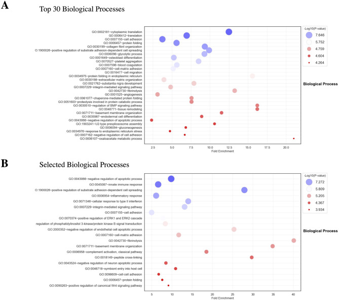
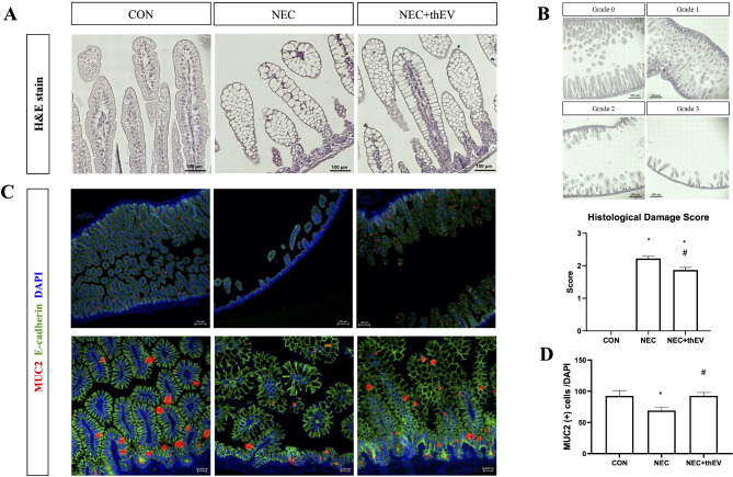
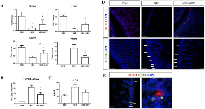
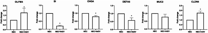
In vitro, compared with naïveMSC-EVs, thMSC-EVs significantly improved cellular viability in HIT-induced IEC-6 cells and reduced pro-inflammatory (IL-1α, IL-1β, TNF-α) but increased anti-inflammatory (TGF-b) cytokine levels in LPS-treated peritoneal macrophages. In vivo, thMSC-EVs significantly attenuated clinical symptoms, reduced intestinal damage, and retained intestinal stem cell markers, showing more significant localization in NEC-induced intestines than in healthy intestines. In NEC mouse-derived organoids, thMSC-EVs significantly increased OLFM4 and claudin-4 expression and reduced stress-related markers such as sucrase-isomaltase, defensin, and chromogranin A. Proteomic analysis revealed that thMSC-EVs were greater enriched in anti-apoptotic, anti-inflammatory, cell adhesion, and Wnt signaling pathways than naïveMSC-EVs.
thMSC-EVs improved cellular viability, reduced apoptosis, attenuated inflammation, and upregulated key intestinal stem cell markers, collectively suggesting their tissue-protective effects and highlighting their potential as a treatment for NEC.
坏死性小肠结肠炎(NEC)是早产儿的一种严重胃肠道疾病,目前尚无特效治疗方法。我们之前证明,凝血酶预处理的间充质基质细胞衍生的细胞外囊泡(thMSC-EVs)可增强对其他新生儿组织损伤的保护作用。因此,本研究旨在评估thMSC-EVs在改良的NEC体外、体内和类器官模型中的治疗潜力。
在体外,比较thMSC-EVs和未处理的间充质基质细胞衍生的细胞外囊泡(naïveMSC-EVs)对高渗、缺血和低温(HIT)应激的IEC-6细胞以及脂多糖(LPS)处理的腹腔巨噬细胞的影响。在体内,通过三次配方奶喂养、口服LPS、缺氧和低温诱导P4小鼠幼崽发生NEC,随后由母鼠过夜照料。连续三天每天腹腔注射2×10个thMSC-EVs,通过宏观、组织学和生化方法评估治疗效果。建立NEC小鼠来源的类器官,以评估thMSC-EVs对成熟肠细胞的作用。采用液相色谱-串联质谱(LC-MS/MS)分析细胞外囊泡蛋白质组学。
在体外,与naïveMSC-EVs相比,thMSC-EVs显著提高了HIT诱导的IEC-6细胞的细胞活力,降低了LPS处理的腹腔巨噬细胞中促炎细胞因子(IL-1α、IL-1β、TNF-α)水平,但提高了抗炎细胞因子(TGF-β)水平。在体内,thMSC-EVs显著减轻临床症状,减少肠道损伤,并保留肠道干细胞标志物,在NEC诱导的肠道中的定位比在健康肠道中更显著。在NEC小鼠来源的类器官中,thMSC-EVs显著增加了OLFM4和claudin-4的表达,并降低了与应激相关的标志物,如蔗糖酶-异麦芽糖酶、防御素和嗜铬粒蛋白A。蛋白质组学分析显示,与naïveMSC-EVs相比,thMSC-EVs在抗凋亡、抗炎、细胞黏附和Wnt信号通路中富集程度更高。
thMSC-EVs提高了细胞活力,减少了细胞凋亡,减轻了炎症,并上调了关键的肠道干细胞标志物,共同表明了它们的组织保护作用,并突出了其作为NEC治疗方法的潜力。