Cheng Kevin, Hii Rachael, Lim Egynne, Yuvaraj Jeremy, Nicholls Stephen J, Dey Damini, Lin Andrew, Wong Dennis T L
Monash Cardiovascular Research Centre, Victorian Heart Institute, Monash University and Monash Health, 631 Blackburn Road, Clayton, Victoria 3168, Australia.
Department of Medicine, Monash University, Wellington Road, Clayton, Victoria 3800, Australia.
Eur Heart J Cardiovasc Imaging. 2025 Apr 30;26(5):784-793. doi: 10.1093/ehjci/jeaf062.
Pericoronary adipose tissue (PCAT) attenuation on coronary computed tomography angiography (CCTA) is an imaging biomarker of coronary inflammation. The natural history of PCAT attenuation remains unknown. High-intensity statin therapy has pleiotropic anti-inflammatory effects. We sought to assess temporal changes in PCAT attenuation in patients with and without statin therapy.
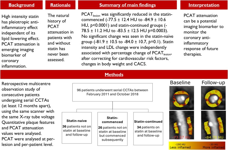
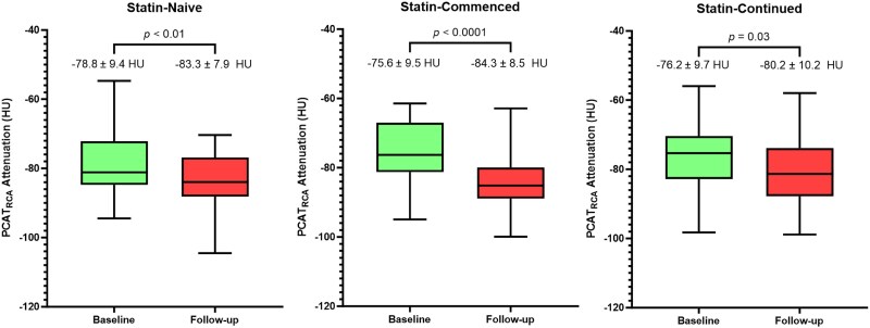
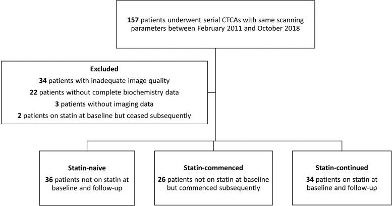
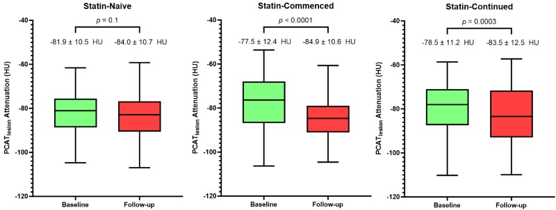
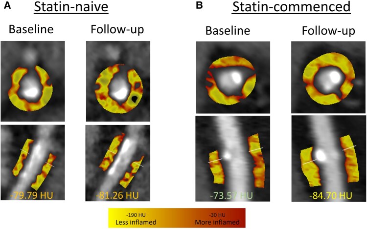
This was a multicentre observational study that included consecutive patients with stable coronary artery disease (CAD) undergoing clinically indicated serial CCTA with identical scan parameters ≥ 12 months apart between May 2013 and July 2022. Using semi-automated software, PCAT attenuation was measured on a per-lesion level (PCATlesion) and per-patient level around the proximal right coronary artery (PCATRCA). Of the 96 patients (57 ± 11 years, 60% male), 34 patients were not on a statin at baseline or follow-up (statin-naive), 26 patients were commenced on a statin after the baseline scan (statin-commenced), and 34 patients were on a statin at both time points (statin-continued). There was no significant difference between the groups for age, sex, body mass index (BMI), and prevalence of traditional cardiovascular risk factors except for dyslipidaemia (25.0% vs. 34.6% vs. 64.7%, P < 0.01 for trend). At a median follow-up of 3.8 years, there was a significant reduction in PCATlesion in the statin-commenced (-79.4 ± 11.7 to -86.5 ± 10 HU, P < 0.001) and the statin-continued (-83.5 ± 8.5 to -90.6 ± 8.5 HU, P = 0.001) groups. Meanwhile, no significant difference in PCATlesion was observed in the statin-naïve group (-84.4 ± 9.7 to -86.6 ± 9.5, P = 0.1). Multivariate analysis showed statin intensity and LDL change to be independently associated with percentage change of PCATlesion, after correcting for cardiovascular risk factors, changes in body weight, and coronary artery calcium score.
Statin therapy was associated with a reduction in PCATlesion, while no significant change in PCATlesion was observed without statin therapy. If validated in larger studies, PCAT attenuation could potentially be used to monitor the response of the coronary arteries to statins and guide treatment.
冠状动脉计算机断层扫描血管造影(CCTA)上的冠状动脉周围脂肪组织(PCAT)衰减是冠状动脉炎症的一种成像生物标志物。PCAT衰减的自然病程尚不清楚。高强度他汀类药物治疗具有多效抗炎作用。我们试图评估接受和未接受他汀类药物治疗患者的PCAT衰减随时间的变化。
这是一项多中心观察性研究,纳入了2013年5月至2022年7月期间连续接受临床指征的系列CCTA检查的稳定型冠状动脉疾病(CAD)患者,扫描参数相同,间隔≥12个月。使用半自动软件,在每个病变水平(PCATlesion)和右冠状动脉近端周围的每位患者水平(PCATRCA)测量PCAT衰减。96例患者(57±11岁,60%为男性)中,34例患者在基线或随访时未服用他汀类药物(未服用他汀类药物组),26例患者在基线扫描后开始服用他汀类药物(开始服用他汀类药物组),34例患者在两个时间点均服用他汀类药物(持续服用他汀类药物组)。除血脂异常外,各组在年龄、性别、体重指数(BMI)和传统心血管危险因素患病率方面无显著差异(分别为25.0%、34.6%和64.7%,趋势P<0.01)。在中位随访3.8年时,开始服用他汀类药物组(从-79.4±11.7至-86.5±10HU,P<0.001)和持续服用他汀类药物组(从-83.5±8.5至-90.6±8.5HU,P=0.001)的PCATlesion显著降低。同时,未服用他汀类药物组的PCATlesion无显著差异(从-84.4±9.7至-86.6±9.5,P=0.1)。多变量分析显示,在校正心血管危险因素、体重变化和冠状动脉钙化积分后,他汀类药物强度和低密度脂蛋白变化与PCATlesion的百分比变化独立相关。
他汀类药物治疗与PCATlesion降低相关,而未接受他汀类药物治疗时PCATlesion无显著变化。如果在更大规模的研究中得到验证,PCAT衰减可能可用于监测冠状动脉对他汀类药物的反应并指导治疗。