Tang Feiyu, Zhu Yongwei, Shen Jia, Yuan Bowen, He Xiang, Tian Yuxi, Weng Liang, Sun Lunquan
Xiangya Cancer Center, Xiangya Hospital, Central South University, Changsha, China.
Key Laboratory of Molecular Radiation Oncology Hunan Province, Changsha, China.
Br J Cancer. 2025 May;132(8):703-715. doi: 10.1038/s41416-025-02968-9. Epub 2025 Mar 12.
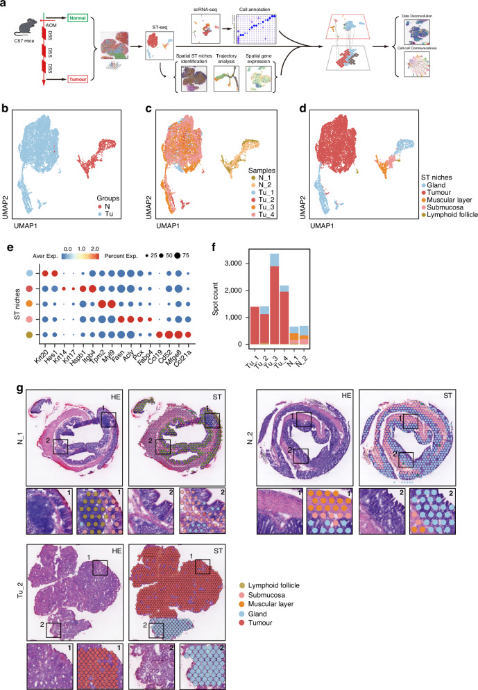
The heterogeneity of tumors significantly impacts on colorectal cancer (CRC) progression. However, the influence of this heterogeneity on the spatial architecture of CRC remains largely unknown.
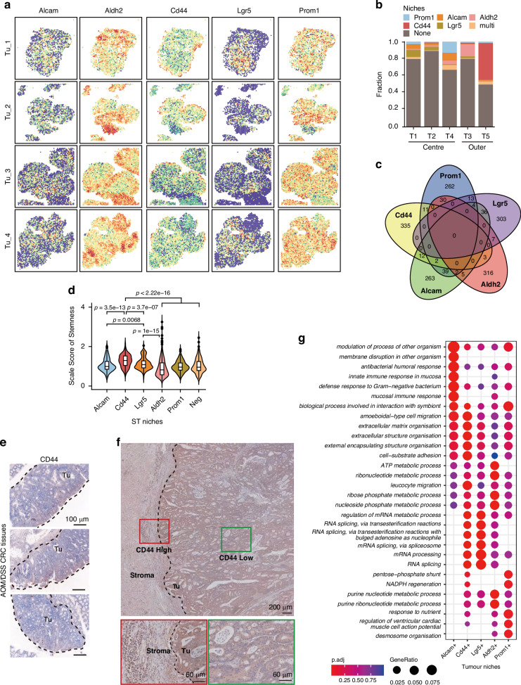
Spatial transcriptomic (ST) analysis of AOM/DSS-induced colorectal cancer (CRC), integrated with single-cell RNA sequencing, generated a comprehensive spatial atlas of CRC. Pseudotime trajectory, stemness evaluation, and cell-cell communication analyses explored how CD44 tumor cells at the leading edge remodel the tumor microenvironment (TME). In vitro experiments and immunofluorescence staining of clinical samples validated pleiotrophin (PTN) signaling in promoting cancer-associated fibroblasts (CAFs) phenotypic transition and CRC progression.
Our findings revealed a distinctive layered ring-like structure within CRC tissues, where CD44 tumor cells exhibiting high stemness were positioned at the tumor's leading edge. Inflammatory CAFs (iCAFs)-like, myofibroblastic CAFs (myCAFs)-like cells and pro-tumorigenic neutrophils primarily located at the tumor edge, in proximity to CD44 tumor cells. CD44 tumor cells then triggered the phenotypic transition of CAFs into iCAF-like and myCAF-like cells through PTN signaling.
Our results provide distinctive insights into how tumor heterogeneity reshapes the TME at the leading edge of tumor, thereby promoting CRC progression.
肿瘤的异质性对结直肠癌(CRC)的进展有显著影响。然而,这种异质性对CRC空间结构的影响在很大程度上仍不清楚。
对AOM/DSS诱导的结直肠癌(CRC)进行空间转录组(ST)分析,并结合单细胞RNA测序,生成了CRC的综合空间图谱。伪时间轨迹、干性评估和细胞间通讯分析探讨了前沿的CD44肿瘤细胞如何重塑肿瘤微环境(TME)。体外实验和临床样本的免疫荧光染色验证了多效生长因子(PTN)信号在促进癌症相关成纤维细胞(CAFs)表型转变和CRC进展中的作用。
我们的研究结果揭示了CRC组织内独特的分层环状结构,其中具有高干性的CD44肿瘤细胞位于肿瘤前沿。炎症性CAFs(iCAFs)样、肌成纤维细胞性CAFs(myCAFs)样细胞和促肿瘤中性粒细胞主要位于肿瘤边缘,靠近CD44肿瘤细胞。然后,CD44肿瘤细胞通过PTN信号触发CAFs向iCAF样和myCAF样细胞的表型转变。
我们的结果为肿瘤异质性如何在肿瘤前沿重塑TME从而促进CRC进展提供了独特的见解。