Shrestha B, Rajan S M, Saunders M, Fawzy A
UWA Dental School, The University of Western Australia, WA, Australia.
Centre for Microscopy, Characterisation and Analysis, The University of Western Australia, WA, Australia.
J Dent Res. 2025 Aug;104(9):983-992. doi: 10.1177/00220345251323869. Epub 2025 Mar 19.
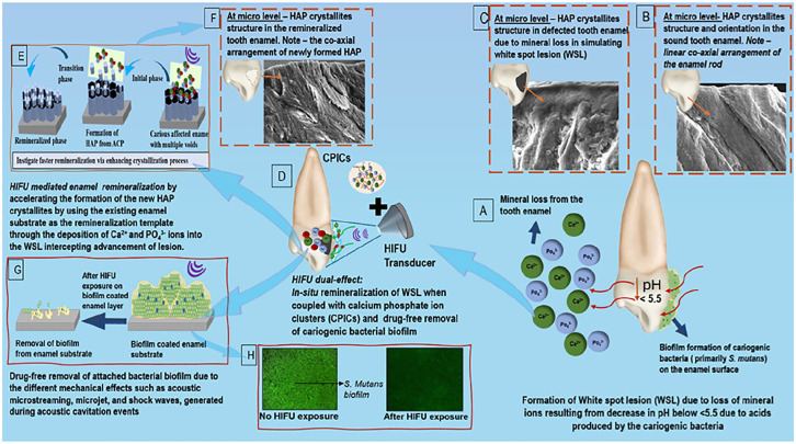
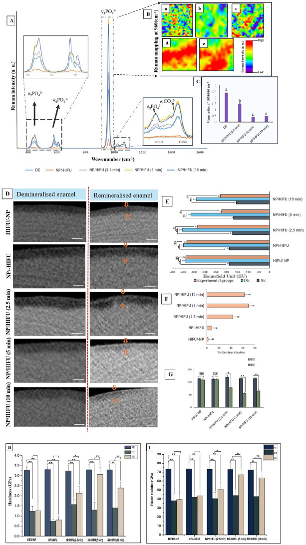
Remineralization is an essential interventional strategy for intercepting enamel white spot lesions (WSLs). Given the limitations of both natural and/or fluoride-mediated repair processes, there is a need to develop novel strategies for repairing enamel WSLs via a minimally invasive approach while restoring the unique ultrastructural integrity and functional properties. Inspired by the unique capability of high-intensity focused ultrasound (HIFU) in facilitating the crystallization process, we propose a novel strategy of employing HIFU for in vitro repair of WSLs through synergizing the crystallization process required for hydroxyapatite (HAP) formation from its precursor (calcium phosphate ion clusters; CPICs). Following CPIC formulation and characterization including the resultant amorphous calcium phosphate (ACP), the effect of HIFU on the ACP-to-HAP transition on the amorphous substrate was investigated using transmission electron microscopy and high-resolution transmission electron microscopy, selected area electron diffraction, and X-ray diffraction (XRD). The results showed profound amorphous-to-crystalline phase transition, within 5- to 30-min HIFU exposure, whereas the long axis of the resultant HAP corresponded with the (002) plane, and a lattice spacing of 0.34 nm indicated a preferred -axis growth direction consistent with the orientation of natural enamel crystallites. For enamel repair, artificial WSLs were created on enamel specimens and then subjected to CPICs, followed by HIFU exposure for 2.5, 5, or 10 min. Scanning electron and atomic force microscopies revealed the decreased surface roughness and the gradual obliteration in the WSL porous structure with continuous linear coaxial arrangement of HAP crystallites filling the prismatic/interprismatic gaps closely resembling sound enamel specifically with 5-min HIFU exposure. Enamel WSL ultrastructural repair was further confirmed from XRD and Raman spectral analyses with the associated regaining of mineral density and nanomechanical properties as reflected from micro-computed tomography (CT) and nanoindentation results, respectively. Micro-CT further validated the subsurface remineralization of WSLs with HIFU exposure. Within the same exposure parameters, HIFU exhibited a potent antibiofilm effect against . This study introduced a new approach for remineralizing enamel WSLs through the potent synergy between HIFU and CPICs.
再矿化是拦截牙釉质白斑病变(WSLs)的一种重要干预策略。鉴于天然和/或氟介导的修复过程存在局限性,需要开发新的策略,通过微创方法修复牙釉质WSLs,同时恢复其独特的超微结构完整性和功能特性。受高强度聚焦超声(HIFU)促进结晶过程的独特能力启发,我们提出了一种新策略,即通过协同羟基磷灰石(HAP)从其前体(磷酸钙离子簇;CPICs)形成所需的结晶过程,利用HIFU对WSLs进行体外修复。在对CPIC进行配方设计和表征(包括所得的无定形磷酸钙(ACP))之后,使用透射电子显微镜、高分辨率透射电子显微镜、选区电子衍射和X射线衍射(XRD)研究了HIFU对无定形基质上ACP向HAP转变的影响。结果表明,在HIFU照射5至30分钟内,发生了从无定形到结晶相的深刻转变,而所得HAP的长轴与(002)面相对应,晶格间距为0.34 nm表明其优先的c轴生长方向与天然牙釉质微晶的取向一致。为了进行牙釉质修复,在牙釉质标本上制造人工WSLs,然后用CPICs处理,随后进行2.5、5或10分钟的HIFU照射。扫描电子显微镜和原子力显微镜显示,表面粗糙度降低,WSLs多孔结构逐渐消失,HAP微晶连续线性同轴排列,紧密填充棱柱/棱柱间隙,特别在5分钟HIFU照射时类似于完好的牙釉质。分别从XRD和拉曼光谱分析进一步证实了牙釉质WSLs的超微结构修复,同时微计算机断层扫描(CT)和纳米压痕结果分别反映了矿物质密度和纳米力学性能的相关恢复。微CT进一步验证了HIFU照射后WSLs的表面下再矿化。在相同的照射参数下,HIFU对表现出强大的抗生物膜作用。本研究通过HIFU与CPICs之间的有效协同作用,引入了一种使牙釉质WSLs再矿化的新方法。