Brügger Melanie, Machahua Carlos, Zumkehr Trix, Cismaru Christiana, Jandrasits Damian, Trüeb Bettina, Ezzat Sara, Oliveira Esteves Blandina I, Dorn Patrick, Marti Thomas M, Zimmer Gert, Thiel Volker, Funke-Chambour Manuela, Alves Marco P
Institute of Virology and Immunology, Bern, Switzerland.
Department of Infectious Diseases and Pathobiology, Vetsuisse Faculty, University of Bern, Bern, Switzerland.
Respir Res. 2025 Mar 24;26(1):112. doi: 10.1186/s12931-025-03190-0.
The coronavirus disease 2019 (COVID-19) outbreak revealed the susceptibility of elderly patients to respiratory virus infections, showing cell senescence or subclinical persistent inflammatory profiles and favoring the development of severe pneumonia.
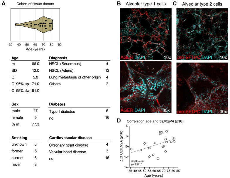
In our study, we evaluated the potential influence of lung aging on the efficiency of replication of influenza A virus (IAV) and severe acute respiratory syndrome coronavirus 2 (SARS-CoV-2), as well as determining the pro-inflammatory and antiviral responses of the distal lung tissue.
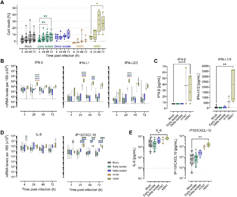
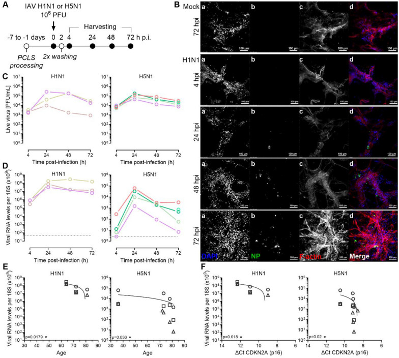
Using precision-cut lung slices (PCLS) from donors of different ages, we found that pandemic H1N1 and avian H5N1 IAV replicated in the lung parenchyma with high efficacy. In contrast to these IAV strains, SARS-CoV-2 Early isolate and Delta variant of concern (VOC) replicated less efficiently in PCLS. Interestingly, both viruses showed reduced replication in PCLS from older compared to younger donors, suggesting that aged lung tissue represents a suboptimal environment for viral replication. Regardless of the age-dependent viral loads, PCLS responded to H5N1 IAV infection by an induction of IL-6 and IP10/CXCL10, both at the mRNA and protein levels, and to H1N1 IAV infection by induction of IP10/CXCL10 mRNA. Finally, while SARS-CoV-2 and H1N1 IAV infection were not causing detectable cell death, H5N1 IAV infection led to more cytotoxicity and induced significant early interferon responses.
In summary, our findings suggest that aged lung tissue might not favor viral dissemination, pointing to a determinant role of dysregulated immune mechanisms in the development of severe disease.
2019年冠状病毒病(COVID-19)疫情揭示了老年患者对呼吸道病毒感染的易感性,表现为细胞衰老或亚临床持续性炎症特征,并有利于重症肺炎的发展。
在我们的研究中,我们评估了肺老化对甲型流感病毒(IAV)和严重急性呼吸综合征冠状病毒2(SARS-CoV-2)复制效率的潜在影响,并确定了远端肺组织的促炎和抗病毒反应。
使用来自不同年龄供体的精密肺切片(PCLS),我们发现大流行H1N1和禽H5N1 IAV在肺实质中高效复制。与这些IAV毒株相反,SARS-CoV-2早期分离株和关注的Delta变异株(VOC)在PCLS中的复制效率较低。有趣的是,与年轻供体相比,两种病毒在老年供体的PCLS中复制均减少,这表明老化的肺组织是病毒复制的次优环境。无论病毒载量是否与年龄相关,PCLS对H5N1 IAV感染的反应是在mRNA和蛋白质水平上诱导IL-6和IP10/CXCL10,对H1N1 IAV感染的反应是诱导IP10/CXCL10 mRNA。最后,虽然SARS-CoV-2和H1N1 IAV感染未导致可检测到的细胞死亡,但H5N1 IAV感染导致更多细胞毒性并诱导显著的早期干扰素反应。
总之,我们的研究结果表明,老化的肺组织可能不利于病毒传播,这表明免疫机制失调在重症疾病发展中起决定性作用。