School of Public Health, Li Ka Shing Faculty of Medicine, The University of Hong Kong, Pokfulam, Special Administrative Region of Hong Kong, China.
Department of Ophthalmology, Li Ka Shing Faculty of Medicine, The University of Hong Kong, Pokfulam, Special Administrative Region of Hong Kong, China.
Lancet Respir Med. 2020 Jul;8(7):687-695. doi: 10.1016/S2213-2600(20)30193-4. Epub 2020 May 7.
Severe acute respiratory syndrome coronavirus 2 (SARS-CoV-2) emerged in December 2019, causing a respiratory disease (coronavirus disease 2019, COVID-19) of varying severity in Wuhan, China, and subsequently leading to a pandemic. The transmissibility and pathogenesis of SARS-CoV-2 remain poorly understood. We evaluate its tissue and cellular tropism in human respiratory tract, conjunctiva, and innate immune responses in comparison with other coronavirus and influenza virus to provide insights into COVID-19 pathogenesis.
We isolated SARS-CoV-2 from a patient with confirmed COVID-19, and compared virus tropism and replication competence with SARS-CoV, Middle East respiratory syndrome-associated coronavirus (MERS-CoV), and 2009 pandemic influenza H1N1 (H1N1pdm) in ex-vivo cultures of human bronchus (n=5) and lung (n=4). We assessed extrapulmonary infection using ex-vivo cultures of human conjunctiva (n=3) and in-vitro cultures of human colorectal adenocarcinoma cell lines. Innate immune responses and angiotensin-converting enzyme 2 expression were investigated in human alveolar epithelial cells and macrophages. In-vitro studies included the highly pathogenic avian influenza H5N1 virus (H5N1) and mock-infected cells as controls.
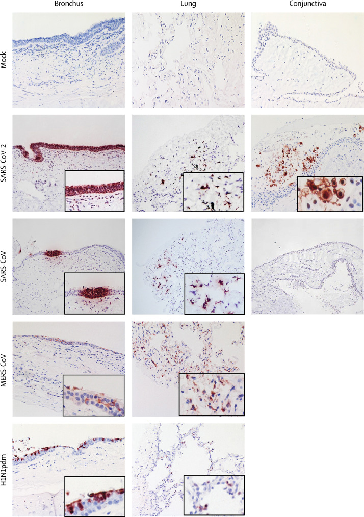
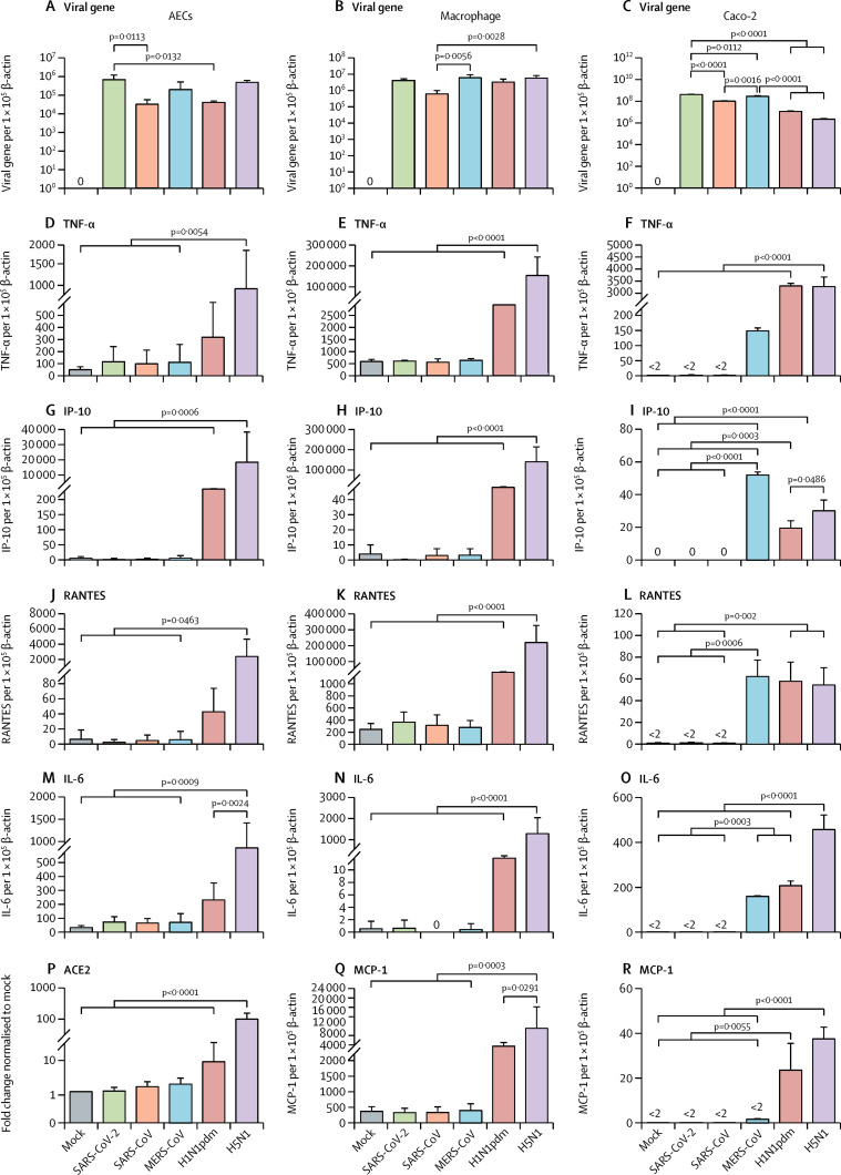
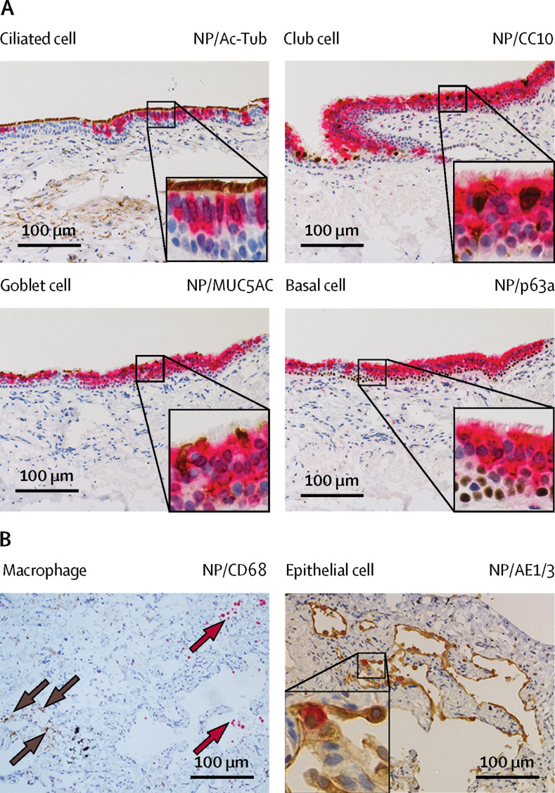
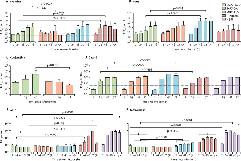
SARS-CoV-2 infected ciliated, mucus-secreting, and club cells of bronchial epithelium, type 1 pneumocytes in the lung, and the conjunctival mucosa. In the bronchus, SARS-CoV-2 replication competence was similar to MERS-CoV, and higher than SARS-CoV, but lower than H1N1pdm. In the lung, SARS-CoV-2 replication was similar to SARS-CoV and H1N1pdm, but was lower than MERS-CoV. In conjunctiva, SARS-CoV-2 replication was greater than SARS-CoV. SARS-CoV-2 was a less potent inducer of proinflammatory cytokines than H5N1, H1N1pdm, or MERS-CoV.
The conjunctival epithelium and conducting airways appear to be potential portals of infection for SARS-CoV-2. Both SARS-CoV and SARS-CoV-2 replicated similarly in the alveolar epithelium; SARS-CoV-2 replicated more extensively in the bronchus than SARS-CoV. These findings provide important insights into the transmissibility and pathogenesis of SARS-CoV-2 infection and differences with other respiratory pathogens.
US National Institute of Allergy and Infectious Diseases, University Grants Committee of Hong Kong Special Administrative Region, China; Health and Medical Research Fund, Food and Health Bureau, Government of Hong Kong Special Administrative Region, China.
严重急性呼吸系统综合症冠状病毒 2 型(SARS-CoV-2)于 2019 年 12 月出现,在中国武汉引发了一种严重程度不同的呼吸道疾病(冠状病毒病 2019,COVID-19),随后导致了大流行。SARS-CoV-2 的传染性和发病机制仍了解甚少。我们将其与其他冠状病毒和流感病毒进行比较,评估其在人类呼吸道、结膜中的组织和细胞趋向性以及先天免疫反应,以深入了解 COVID-19 的发病机制。
我们从一名确诊 COVID-19 的患者中分离出 SARS-CoV-2,并在体外培养的人类支气管(n=5)和肺(n=4)中比较病毒趋向性和复制能力与 SARS-CoV、中东呼吸综合征相关冠状病毒(MERS-CoV)和 2009 年大流行性流感 H1N1(H1N1pdm)。我们使用体外培养的人结膜(n=3)和人结直肠腺癌细胞系的体外培养来评估肺外感染。我们在人肺泡上皮细胞和巨噬细胞中研究了先天免疫反应和血管紧张素转换酶 2 的表达。体外研究包括高致病性禽流感 H5N1 病毒(H5N1)和模拟感染细胞作为对照。
SARS-CoV-2 感染了支气管上皮的纤毛、分泌黏液的细胞和分泌黏液的细胞,以及肺部的 1 型肺泡细胞和结膜黏膜。在支气管中,SARS-CoV-2 的复制能力与 MERS-CoV 相似,高于 SARS-CoV,但低于 H1N1pdm。在肺部,SARS-CoV-2 的复制与 SARS-CoV 和 H1N1pdm 相似,但低于 MERS-CoV。在结膜中,SARS-CoV-2 的复制能力大于 SARS-CoV。SARS-CoV-2 诱导促炎细胞因子的能力低于 H5N1、H1N1pdm 或 MERS-CoV。
结膜上皮和传导气道似乎是 SARS-CoV-2 的潜在感染门户。SARS-CoV 和 SARS-CoV-2 都在肺泡上皮中复制;SARS-CoV-2 在支气管中的复制比 SARS-CoV 更广泛。这些发现为 SARS-CoV-2 感染的传染性和发病机制以及与其他呼吸道病原体的差异提供了重要的见解。
美国国立过敏和传染病研究所、香港特别行政区大学教育资助委员会、中国;香港特别行政区政府食物及卫生局卫生及医疗研究基金。