Frangos Sophia M, Damrich Sebastian, Gueiber Daniele, Sanchez Cecilia P, Wiedemann Philipp, Schwarz Ulrich S, Hamprecht Fred A, Lanzer Michael
Heidelberg University, Medical Faculty, University Hospital Heidelberg, Center for Infectious Diseases, Parasitology, Im Neuenheimer Feld 324, Heidelberg, Germany.
Heidelberg University, Interdisciplinary Center for Scientific Computing (IWR), Im Neuenheimer Feld 205, Heidelberg, Germany.
Commun Biol. 2025 Mar 25;8(1):487. doi: 10.1038/s42003-025-07894-3.
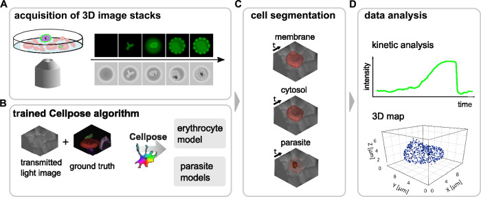
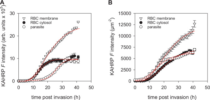
Continuous high-resolution imaging of the disease-mediating blood stages of the human malaria parasite Plasmodium falciparum faces challenges due to photosensitivity, small parasite size, and the anisotropy and large refractive index of host erythrocytes. Previous studies often relied on snapshot galleries from multiple cells, limiting the investigation of dynamic cellular processes. We present a workflow enabling continuous, single-cell monitoring of live parasites throughout the 48-hour intraerythrocytic life cycle with high spatial and temporal resolution. This approach integrates label-free, three-dimensional differential interference contrast and fluorescence imaging using an Airyscan microscope, automated cell segmentation through pre-trained deep-learning algorithms, and 3D rendering for visualization and time-resolved analyses. As a proof of concept, we applied this workflow to study knob-associated histidine-rich protein (KAHRP) export into the erythrocyte compartment and its clustering beneath the plasma membrane. Our methodology opens avenues for in-depth exploration of dynamic cellular processes in malaria parasites, providing a valuable tool for further investigations.
由于疟原虫的光敏性、寄生虫尺寸小以及宿主红细胞的各向异性和大折射率,对人类疟原虫恶性疟原虫介导疾病的血液阶段进行连续高分辨率成像面临挑战。以往的研究通常依赖于多个细胞的快照图库,限制了对动态细胞过程的研究。我们提出了一种工作流程,能够在48小时的红细胞内生命周期中以高空间和时间分辨率对活寄生虫进行连续的单细胞监测。这种方法集成了使用Airyscan显微镜的无标记三维微分干涉对比和荧光成像、通过预训练的深度学习算法进行自动细胞分割以及用于可视化和时间分辨分析的三维渲染。作为概念验证,我们应用此工作流程研究富含组氨酸的旋钮相关蛋白(KAHRP)输出到红细胞区室及其在质膜下的聚集。我们的方法为深入探索疟原虫的动态细胞过程开辟了道路,为进一步研究提供了有价值的工具。