• 文献检索
  • 文档翻译
  • 深度研究
  • 学术资讯
  • Suppr Zotero 插件Zotero 插件
  • 邀请有礼
  • 套餐&价格
  • 历史记录
应用&插件
Suppr Zotero 插件Zotero 插件浏览器插件Mac 客户端Windows 客户端微信小程序
定价
高级版会员购买积分包购买API积分包
服务
文献检索文档翻译深度研究API 文档MCP 服务
关于我们
关于 Suppr公司介绍联系我们用户协议隐私条款
关注我们

Suppr 超能文献

核心技术专利:CN118964589B侵权必究
粤ICP备2023148730 号-1Suppr @ 2026

文献检索

告别复杂PubMed语法,用中文像聊天一样搜索,搜遍4000万医学文献。AI智能推荐,让科研检索更轻松。

立即免费搜索

文件翻译

保留排版,准确专业,支持PDF/Word/PPT等文件格式,支持 12+语言互译。

免费翻译文档

深度研究

AI帮你快速写综述,25分钟生成高质量综述,智能提取关键信息,辅助科研写作。

立即免费体验

肾小球基底膜连续性中断。无细胞性肾小球的扫描电子显微镜研究。

Glomerular basement membrane discontinuities. Scanning electron microscopic study of acellular glomeruli.

作者信息

Bonsib S M

出版信息

Am J Pathol. 1985 Jun;119(3):357-60.

PMID:4014431
原文链接:https://pmc.ncbi.nlm.nih.gov/articles/PMC1888009/
Abstract

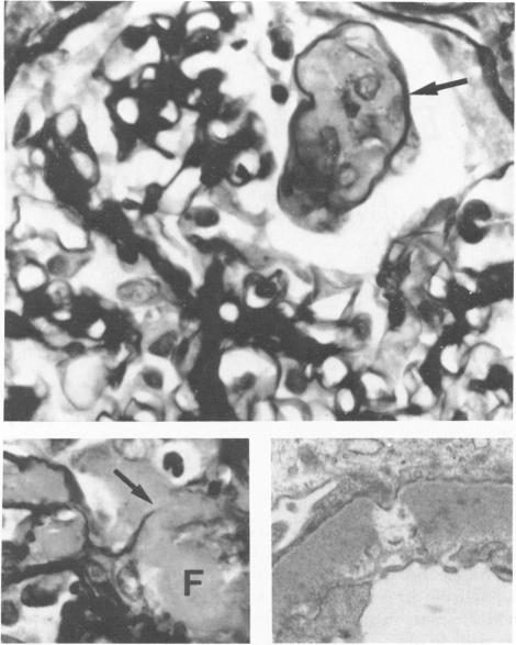
Glomerulonephritides which develop necrotizing and crescentic lesions usually have glomerular basement membrane (GBM) disruptions when carefully examined by light microscopy or transmission electron microscopy. Despite numerous excellent and detailed ultrastructural investigations of GBM discontinuities, a complete appreciation of their actual number, appearance, and distribution within a glomerulus has been difficult to achieve by reconstruction of two-dimensional light or transmission electron microscopic images. Selective removal of podocytes by a sequence of lytic and solubilization procedures has been developed which exposes any structural alteration of the GBM to direct examination by scanning electron microscopy. A case of idiopathic, immune-complex-negative, focal-segmental necrotizing glomerulonephritis has been studied by this technique, permitting three-dimensional visualization of the GBM defects which result in free communication between the vascular and urinary spaces. These disruptions were distinctive by their frequency within an affected lobule, variable size, and sharply demarcated edges. Application of this technique to human renal biopsies is capable of enhancing our understanding of the morphologic alterations occurring in human glomerulonephritis.

摘要

通常,发展为坏死性和新月体性病变的肾小球肾炎,经光学显微镜或透射电子显微镜仔细检查时,肾小球基底膜(GBM)会出现中断。尽管对GBM连续性进行了大量出色且详细的超微结构研究,但通过二维光学或透射电子显微镜图像重建,很难全面了解其在肾小球内的实际数量、外观和分布。已开发出一系列溶解和增溶程序来选择性去除足细胞,从而使GBM的任何结构改变都能通过扫描电子显微镜直接检查。通过该技术对一例特发性、免疫复合物阴性、局灶节段性坏死性肾小球肾炎病例进行了研究,从而能够三维可视化导致血管腔和尿腔自由连通的GBM缺陷。这些中断在受影响的小叶内的频率、大小各异以及边缘清晰可辨,具有独特性。将该技术应用于人类肾活检,能够增进我们对人类肾小球肾炎中发生的形态学改变的理解。

https://cdn.ncbi.nlm.nih.gov/pmc/blobs/2a78/1888009/2c8f3be9116b/amjpathol00171-0013-a.jpg
https://cdn.ncbi.nlm.nih.gov/pmc/blobs/2a78/1888009/852911830d36/amjpathol00171-0012-a.jpg
https://cdn.ncbi.nlm.nih.gov/pmc/blobs/2a78/1888009/2c8f3be9116b/amjpathol00171-0013-a.jpg
https://cdn.ncbi.nlm.nih.gov/pmc/blobs/2a78/1888009/852911830d36/amjpathol00171-0012-a.jpg
https://cdn.ncbi.nlm.nih.gov/pmc/blobs/2a78/1888009/2c8f3be9116b/amjpathol00171-0013-a.jpg

相似文献

1
Glomerular basement membrane discontinuities. Scanning electron microscopic study of acellular glomeruli.肾小球基底膜连续性中断。无细胞性肾小球的扫描电子显微镜研究。
Am J Pathol. 1985 Jun;119(3):357-60.
2
SEM studies of acellular glomerular basement membrane in human diabetic glomerulopathy.人糖尿病肾小球病中无细胞肾小球基底膜的扫描电子显微镜研究
Anat Rec. 1986 Nov;216(3):349-58. doi: 10.1002/ar.1092160303.
3
Ultrastructural change of the glomerular basement membrane in rats with Heymann nephritis revealed by ultrahigh resolution scanning electron microscopy.超高分辨率扫描电子显微镜揭示的海曼肾炎大鼠肾小球基底膜的超微结构变化
J Pathol. 1996 May;179(1):112-20. doi: 10.1002/(SICI)1096-9896(199605)179:1<112::AID-PATH542>3.0.CO;2-Q.
4
Three-dimensional ultrastructural changes of acellular glomerular basement membrane in various types of human glomerulonephritis.
Nephron. 1989;53(1):9-17. doi: 10.1159/000185694.
5
The evolution of crescentic nephritis and alveolar haemorrhage following induction of autoimmunity to glomerular basement membrane in an experimental model of Goodpasture's disease.在古德帕斯彻氏病实验模型中,诱导针对肾小球基底膜的自身免疫后新月体性肾炎和肺泡出血的演变。
J Pathol. 2003 May;200(1):118-29. doi: 10.1002/path.1336.
6
Segmental subepithelial deposits in primary glomerulonephritis: scanning electron microscopic examination of acellular glomeruli.原发性肾小球肾炎中的节段性上皮下沉积物:无细胞肾小球的扫描电子显微镜检查
Hum Pathol. 1985 Nov;16(11):1115-21. doi: 10.1016/s0046-8177(85)80179-9.
7
Indentations of the glomerular basement membrane in renal diseases. A light and electron microscopic study on ultrathin serial sections.肾脏疾病中肾小球基底膜的压迹。对超薄连续切片的光镜和电镜研究。
Lab Invest. 1976 Oct;35(4):327-36.
8
Scanning electron microscopy of acellular glomeruli in human glomerulonephritis: application of a technique.
Ultrastruct Pathol. 1984;7(2-3):215-7. doi: 10.3109/01913128409141479.
9
Scanning electron microscopy of the acellular glomerular basement membranes in idiopathic membranous glomerulopathy.
Lab Invest. 1986 Jan;54(1):84-92.
10
Chronic Masugi nephritis in the rat. An electron microscopic study on evolution and consequences of glomerular capsular adhesions.大鼠慢性马杉肾炎。肾小球囊粘连演变及后果的电子显微镜研究。
Acta Pathol Jpn. 1982 Mar;32(2):231-42.

引用本文的文献

1
Early and late scanning electron microscopy findings in diabetic kidney disease.糖尿病肾病的早期和晚期扫描电子显微镜观察结果。
Sci Rep. 2018 Mar 20;8(1):4909. doi: 10.1038/s41598-018-23244-2.
2
Neutrophil Extracellular Trap-Related Extracellular Histones Cause Vascular Necrosis in Severe GN.中性粒细胞胞外诱捕网相关的细胞外组蛋白在重症肾小球肾炎中导致血管坏死。
J Am Soc Nephrol. 2015 Oct;26(10):2399-413. doi: 10.1681/ASN.2014070673. Epub 2015 Feb 2.
3
Henoch-Schönlein purpura nephritis in children.儿童过敏性紫癜性肾炎。

本文引用的文献

1
The structural basis of proteinuria in man; electron microscopic studies of renal biopsy specimens from patients with lipid nephrosis, amyloidosis, and subacute and chronic glomerulonephritis.人类蛋白尿的结构基础;对脂性肾病、淀粉样变性以及亚急性和慢性肾小球肾炎患者肾活检标本的电子显微镜研究。
Am J Pathol. 1959 Jan-Feb;35(1):47-73.
2
Idiopathic rapidly progressive glomerulonephritis.特发性快速进展性肾小球肾炎
Am J Nephrol. 1982;2(2):57-69. doi: 10.1159/000166586.
3
Scanning electron microscopy of acellular glomeruli in human glomerulonephritis: application of a technique.
Nat Rev Nephrol. 2014 Oct;10(10):563-73. doi: 10.1038/nrneph.2014.126. Epub 2014 Jul 29.
4
Danger control programs cause tissue injury and remodeling.危险控制程序会导致组织损伤和重塑。
Int J Mol Sci. 2013 May 28;14(6):11319-46. doi: 10.3390/ijms140611319.
5
Four danger response programs determine glomerular and tubulointerstitial kidney pathology: clotting, inflammation, epithelial and mesenchymal healing.四种危险反应程序决定肾小球和肾小管间质肾脏病理:凝血、炎症、上皮和间充质愈合。
Organogenesis. 2012 Apr-Jun;8(2):29-40. doi: 10.4161/org.20342. Epub 2012 Apr 1.
6
[Rapidly progressive glomerulonephritis:classification, pathogenesis and clinical management].[快速进展性肾小球肾炎:分类、发病机制及临床管理]
Internist (Berl). 2003 Sep;44(9):1107-19. doi: 10.1007/s00108-003-1025-2.
7
Scanning electron microscopy of cellular and acellular glomeruli of male dogs affected with Samoyed hereditary glomerulopathy and a carrier female.患有萨摩耶遗传性肾小球病的雄性犬以及一只携带致病基因的雌性犬的细胞性和无细胞性肾小球的扫描电子显微镜检查
Can J Vet Res. 1987 Oct;51(4):475-80.
8
Alterations of glomerular basement membrane relevant to haematuria.
Virchows Arch A Pathol Anat Histopathol. 1988;413(2):159-65. doi: 10.1007/BF00749678.
9
Three-dimensional studies of acellular glomerular basement membranes in dense-deposit disease.致密物沉积病中无细胞肾小球基底膜的三维研究
Virchows Arch A Pathol Anat Histopathol. 1986;409(5):595-607. doi: 10.1007/BF00713427.
Ultrastruct Pathol. 1984;7(2-3):215-7. doi: 10.3109/01913128409141479.
4
Discontinuities (gaps) of the glomerular capillary wall and basement membrane in renal diseases.肾脏疾病中肾小球毛细血管壁和基底膜的连续性中断(间隙)。
Lab Invest. 1973 Feb;28(2):149-69.
5
The morphogenesis of glomerular crescents in rapidly progressive glomerulonephritis.快速进行性肾小球肾炎中肾小球新月体的形态发生
Kidney Int. 1974 Jan;5(1):47-56. doi: 10.1038/ki.1974.6.
6
Ultrastructural demonstration of injury and perforation of glomerular capillary basement membrane in acute proliferative glomerulonephritis.急性增生性肾小球肾炎肾小球毛细血管基底膜损伤与穿孔的超微结构显示
Am J Pathol. 1969 Aug;56(2):251-65.
7
Glomerular ultrastructure in extracapillary glomerulonephritis.毛细血管外肾小球肾炎的肾小球超微结构
Acta Pathol Microbiol Scand Suppl. 1974;Suppl 249:29-54.
8
Structure and development of the glomerular crescent.肾小球新月体的结构与发育
Am J Pathol. 1973 Sep;72(3):349-68.