• 文献检索
  • 文档翻译
  • 深度研究
  • 学术资讯
  • Suppr Zotero 插件Zotero 插件
  • 邀请有礼
  • 套餐&价格
  • 历史记录
应用&插件
Suppr Zotero 插件Zotero 插件浏览器插件Mac 客户端Windows 客户端微信小程序
定价
高级版会员购买积分包购买API积分包
服务
文献检索文档翻译深度研究API 文档MCP 服务
关于我们
关于 Suppr公司介绍联系我们用户协议隐私条款
关注我们

Suppr 超能文献

核心技术专利:CN118964589B侵权必究
粤ICP备2023148730 号-1Suppr @ 2026

文献检索

告别复杂PubMed语法,用中文像聊天一样搜索,搜遍4000万医学文献。AI智能推荐,让科研检索更轻松。

立即免费搜索

文件翻译

保留排版,准确专业,支持PDF/Word/PPT等文件格式,支持 12+语言互译。

免费翻译文档

深度研究

AI帮你快速写综述,25分钟生成高质量综述,智能提取关键信息,辅助科研写作。

立即免费体验

不同临床阶段的Leber遗传性视神经病变中神经节细胞和神经纤维层的改变。

Alterations in ganglion cell and nerve fiber layer in Leber hereditary optic neuropathy across clinical stages.

作者信息

Oeverhaus Michael, Knetsch Mareile, Chen Ying, Jabbarli Leyla, Nolden Carmen, Eckstein Anja, Bechrakis Nikolaos E, Rating Philipp

机构信息

Department of Ophthalmology, University Hospital Essen, Essen, Germany.

Medical Practice Dres. Oeverhaus, Rietberg, Germany.

出版信息

BMC Ophthalmol. 2025 Apr 8;25(1):183. doi: 10.1186/s12886-025-03991-3.

DOI:10.1186/s12886-025-03991-3
PMID:40200205
原文链接:https://pmc.ncbi.nlm.nih.gov/articles/PMC11977937/
Abstract

PURPOSE

LHON leads to gradual, painless, and permanent vision loss in both eyes, often associated with central scotomas. As the condition progresses, there is a decline in visual function, accompanied by noticeable structural alterations. This study focused on evaluating the clinical characteristics of patients with differing LHON stages, with a specific emphasis on optical coherence tomography (OCT) imaging results.

METHODS

This analysis included 22 individuals with LHON. Patients underwent thorough clinical ophthalmologic assessments, including SD-OCT, Visual evoked potentials, and perimetry. When LHON was suspected, blood samples were obtained to test for the three major mitochondrial mutations (G1178A, T14484C, G3460A), with further sequencing to identify additional known mutations. The data were subsequently examined through descriptive statistical methods.

RESULTS

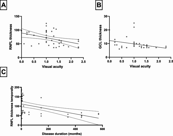
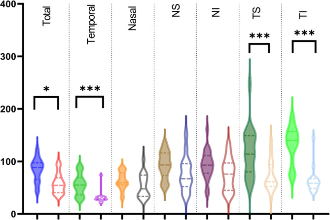
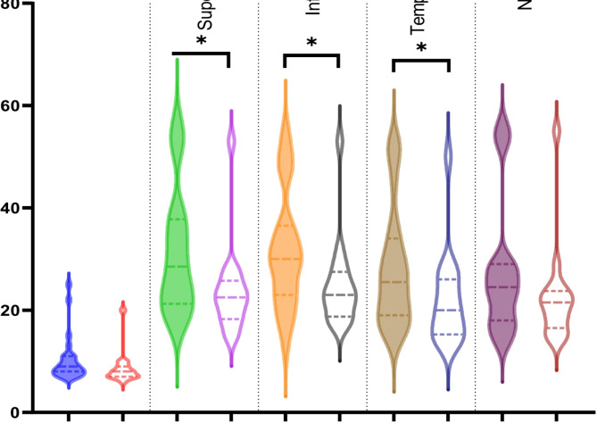
The clinical characteristics of 22 individuals (median age 33, range 9-68) were examined. All participants carried a mutation linked to LHON. The most prevalent mutation was G11778A (55%), followed by G3460A (23%), T14484C (14%), with one instance each of the rare G13042A and C3461T mutations. Fourteen participants experienced acute vision loss (average duration: 5.2 ± 5 months), while eight had chronic LHON. There was no significant difference in visual acuity (VA, logMAR) between the two groups (0.9 vs. 0.9, p = 0.91). However, chronic patients exhibited significantly reduced the retinal nerve fiber layer (RNFL), especially in the temporal region (32 μm vs. 56 μm, p < 0.0001), but not in the nasal region. Ganglion cell layer (GCL) thickness was also notably thinner in the temporal area for chronic patients compared to those with acute LHON (22 μm vs. 28 μm, p = 0.04). Linear regression analysis showed correlations between RNFL and GCL and visual acuity (R² = 0.18, p = 0.007 and R² = 0.1, p = 0.05).

CONCLUSION

In our analysis, we observed an unusual pattern in the genetic mutations, with G3460A being the second most frequent, rather than T14484C, which may be attributed to the limited sample size. 14 patients experienced acute or subacute vision loss, while eight were assessed for chronic disease. Those with chronic LHON demonstrated significantly thinner GCL and RNFL. These results underscore the importance of accelerating both diagnosis and treatment to facilitate prompt intervention for patients.

摘要

目的

Leber遗传性视神经病变(LHON)会导致双眼逐渐、无痛且永久性的视力丧失,常伴有中心暗点。随着病情发展,视觉功能会下降,并伴有明显的结构改变。本研究着重评估不同LHON阶段患者的临床特征,特别关注光学相干断层扫描(OCT)成像结果。

方法

该分析纳入了22例LHON患者。患者接受了全面的临床眼科评估,包括频域光学相干断层扫描(SD - OCT)、视觉诱发电位和视野检查。当怀疑为LHON时,采集血样检测三种主要的线粒体突变(G1178A、T14484C、G3460A),并进一步测序以确定其他已知突变。随后通过描述性统计方法对数据进行检查。

结果

对22例患者(中位年龄33岁,范围9 - 68岁)的临床特征进行了检查。所有参与者都携带与LHON相关的突变。最常见的突变是G11778A(55%),其次是G3460A(23%)、T14484C(14%),还有罕见的G13042A和C3461T突变各1例。14名参与者经历了急性视力丧失(平均持续时间:5.2±5个月),而8例为慢性LHON。两组之间的视力(VA,对数最小分辨角视力)无显著差异(0.9对0.9,p = 0.91)。然而,慢性病患者的视网膜神经纤维层(RNFL)明显变薄,尤其是在颞侧区域(32μm对56μm,p < 0.0001),但鼻侧区域没有。与急性LHON患者相比,慢性病患者颞侧区域的神经节细胞层(GCL)厚度也明显更薄(22μm对28μm,p = 0.04)。线性回归分析显示RNFL和GCL与视力之间存在相关性(R² = 0.18,p = 0.007和R² = 0.1,p = 0.05)。

结论

在我们的分析中,我们观察到基因突变存在异常模式,G3460A是第二常见的,而非T14484C,这可能归因于样本量有限。14例患者经历了急性或亚急性视力丧失,8例被评估为慢性病。慢性LHON患者的GCL和RNFL明显更薄。这些结果强调了加快诊断和治疗以促进对患者及时干预的重要性。

https://cdn.ncbi.nlm.nih.gov/pmc/blobs/ba24/11977937/8b9c6cfd47df/12886_2025_3991_Fig6_HTML.jpg
https://cdn.ncbi.nlm.nih.gov/pmc/blobs/ba24/11977937/5b325f868bba/12886_2025_3991_Fig1_HTML.jpg
https://cdn.ncbi.nlm.nih.gov/pmc/blobs/ba24/11977937/643159d86aa7/12886_2025_3991_Fig3_HTML.jpg
https://cdn.ncbi.nlm.nih.gov/pmc/blobs/ba24/11977937/f55a5e99eaeb/12886_2025_3991_Fig5_HTML.jpg
https://cdn.ncbi.nlm.nih.gov/pmc/blobs/ba24/11977937/8b9c6cfd47df/12886_2025_3991_Fig6_HTML.jpg

相似文献

1
Alterations in ganglion cell and nerve fiber layer in Leber hereditary optic neuropathy across clinical stages.不同临床阶段的Leber遗传性视神经病变中神经节细胞和神经纤维层的改变。
BMC Ophthalmol. 2025 Apr 8;25(1):183. doi: 10.1186/s12886-025-03991-3.
2
Clinical follow-up investigation on thickness changes in the peripapillary retinal nerve fibre layer of patients with Leber hereditary optic neuropathy.Leber 遗传性视神经病变患者视盘周围视网膜神经纤维层厚度变化的临床随访研究。
BMC Ophthalmol. 2024 Aug 15;24(1):348. doi: 10.1186/s12886-024-03625-0.
3
Natural History of Conversion of Leber's Hereditary Optic Neuropathy: A Prospective Case Series.Leber 遗传性视神经病变的自然病程:一项前瞻性病例系列研究。
Ophthalmology. 2017 Jun;124(6):843-850. doi: 10.1016/j.ophtha.2017.01.002. Epub 2017 Feb 10.
4
Optical Coherence Tomography of the Retinal Ganglion Cell Complex in Leber's Hereditary Optic Neuropathy and Dominant Optic Atrophy.Leber 遗传性视神经病变和显性视神经萎缩的视网膜神经节细胞复合体的光相干断层扫描。
Curr Eye Res. 2019 Jun;44(6):638-644. doi: 10.1080/02713683.2019.1567792. Epub 2019 Feb 4.
5
Leber hereditary optic neuropathy gene therapy clinical trial recruitment: year 1.Leber遗传性视神经病变基因治疗临床试验招募:第1年。
Arch Ophthalmol. 2010 Sep;128(9):1129-35. doi: 10.1001/archophthalmol.2010.201.
6
Macular Retinal Sublayer Thicknesses in G11778A Leber Hereditary Optic Neuropathy.G11778A 型 Leber 遗传性视神经病变的黄斑视网膜各亚层厚度
Ophthalmic Surg Lasers Imaging Retina. 2016 Sep 1;47(9):802-10. doi: 10.3928/23258160-20160901-02.
7
Retinal Ganglion Cell and Inner Plexiform Layer Loss Correlate with Visual Acuity Loss in LHON: A Longitudinal, Segmentation OCT Analysis.视网膜神经节细胞和内网状层丢失与Leber遗传性视神经病变的视力丧失相关:一项纵向、分割光学相干断层扫描分析
Invest Ophthalmol Vis Sci. 2016 Jul 1;57(8):3872-83. doi: 10.1167/iovs.15-17328.
8
Retinal nerve fiber layer thickness variability in Leber hereditary optic neuropathy carriers.Leber遗传性视神经病变携带者的视网膜神经纤维层厚度变异性
Eur J Ophthalmol. 2012 Nov-Dec;22(6):985-91. doi: 10.5301/ejo.5000154. Epub 2012 May 4.
9
Trial end points and natural history in patients with G11778A Leber hereditary optic neuropathy : preparation for gene therapy clinical trial.G11778A 型莱伯遗传性视神经病变患者的临床试验终点和自然史:基因治疗临床试验的准备。
JAMA Ophthalmol. 2014 Apr 1;132(4):428-36. doi: 10.1001/jamaophthalmol.2013.7971.
10
Visual function in chronic Leber's hereditary optic neuropathy during idebenone treatment initiated 5 to 50 years after onset.发病后 5 至 50 年开始用艾地苯醌治疗莱伯遗传性视神经病变的慢性期的视觉功能。
Graefes Arch Clin Exp Ophthalmol. 2019 Dec;257(12):2751-2757. doi: 10.1007/s00417-019-04444-6. Epub 2019 Sep 3.

本文引用的文献

1
Variability of relationship between inner-retinal structural changes and visual dysfunction in optic neuropathy.视神经病变中外层视网膜结构变化与视觉功能障碍关系的可变性。
Sci Rep. 2024 May 27;14(1):12069. doi: 10.1038/s41598-024-62704-w.
2
Gene therapy for mitochondrial disorders.线粒体疾病的基因治疗。
J Inherit Metab Dis. 2024 Jan;47(1):145-175. doi: 10.1002/jimd.12699. Epub 2024 Jan 3.
3
Mitochondrial optic neuropathies.线粒体视神经病变
Handb Clin Neurol. 2023;194:23-42. doi: 10.1016/B978-0-12-821751-1.00010-5.
4
The Relative Preservation of the Central Retinal Layers in Leber Hereditary Optic Neuropathy.莱伯遗传性视神经病变中视网膜中央层的相对保留情况
J Clin Med. 2022 Oct 13;11(20):6045. doi: 10.3390/jcm11206045.
5
Long-Term Follow-Up After Unilateral Intravitreal Gene Therapy for Leber Hereditary Optic Neuropathy: The RESTORE Study.单侧玻璃体内基因治疗莱伯遗传性视神经病变的长期随访:RESTORE 研究。
J Neuroophthalmol. 2021 Sep 1;41(3):309-315. doi: 10.1097/WNO.0000000000001367.
6
Safety of Intravitreal Gene Therapy for Treatment of Subjects with Leber Hereditary Optic Neuropathy due to Mutations in the Mitochondrial ND4 Gene: The REVEAL Study.治疗由线粒体 ND4 基因突变引起的 Leber 遗传性视神经病变患者的玻璃体内基因治疗的安全性:REVEAL 研究。
BioDrugs. 2021 Mar;35(2):201-214. doi: 10.1007/s40259-021-00468-9. Epub 2021 Feb 10.
7
Real-World Clinical Experience With Idebenone in the Treatment of Leber Hereditary Optic Neuropathy.依地酸-beta-羟乙酯治疗莱伯遗传性视神经病变的真实世界临床经验。
J Neuroophthalmol. 2020 Dec;40(4):558-565. doi: 10.1097/WNO.0000000000001023.
8
Demographics of a Large International Population of Patients Affected by Leber's Hereditary Optic Neuropathy.莱伯遗传性视神经病变大国际患者人群的人口统计学。
Ophthalmology. 2020 May;127(5):679-688. doi: 10.1016/j.ophtha.2019.11.014. Epub 2019 Nov 25.
9
Choroidal thickness and the retinal ganglion cell complex in chronic Leber's hereditary optic neuropathy: a prospective study using swept-source optical coherence tomography.慢性莱伯遗传性视神经病变脉络膜厚度和视网膜神经节细胞复合体:应用扫频源光学相干断层扫描的前瞻性研究。
Eye (Lond). 2020 Sep;34(9):1624-1630. doi: 10.1038/s41433-019-0695-5. Epub 2019 Dec 5.
10
Optical Coherence Tomography of the Retinal Ganglion Cell Complex in Leber's Hereditary Optic Neuropathy and Dominant Optic Atrophy.Leber 遗传性视神经病变和显性视神经萎缩的视网膜神经节细胞复合体的光相干断层扫描。
Curr Eye Res. 2019 Jun;44(6):638-644. doi: 10.1080/02713683.2019.1567792. Epub 2019 Feb 4.