• 文献检索
  • 文档翻译
  • 深度研究
  • 学术资讯
  • Suppr Zotero 插件Zotero 插件
  • 邀请有礼
  • 套餐&价格
  • 历史记录
应用&插件
Suppr Zotero 插件Zotero 插件浏览器插件Mac 客户端Windows 客户端微信小程序
定价
高级版会员购买积分包购买API积分包
服务
文献检索文档翻译深度研究API 文档MCP 服务
关于我们
关于 Suppr公司介绍联系我们用户协议隐私条款
关注我们

Suppr 超能文献

核心技术专利:CN118964589B侵权必究
粤ICP备2023148730 号-1Suppr @ 2026

文献检索

告别复杂PubMed语法,用中文像聊天一样搜索,搜遍4000万医学文献。AI智能推荐,让科研检索更轻松。

立即免费搜索

文件翻译

保留排版,准确专业,支持PDF/Word/PPT等文件格式,支持 12+语言互译。

免费翻译文档

深度研究

AI帮你快速写综述,25分钟生成高质量综述,智能提取关键信息,辅助科研写作。

立即免费体验

人工智能与患者护理的一致性:一项关于乳腺密度评估的大规模纵向研究

Artificial intelligence and consistency in patient care: a large-scale longitudinal study of mammographic density assessment.

作者信息

Holley Susan O, Cardoza Daniel, Matthews Thomas P, Tibatemwa Elisha E, Morales Hoil Rodrigo, Toriola Adetunji T, Gastounioti Aimilia

机构信息

Onsite Women's Health, Nashville, TN 37203, United States.

Whiterabbit.ai, Redwood City, CA 94065, United States.

出版信息

BJR Artif Intell. 2025 Mar 3;2(1):ubaf004. doi: 10.1093/bjrai/ubaf004. eCollection 2025 Jan.

DOI:10.1093/bjrai/ubaf004
PMID:40201185
原文链接:https://pmc.ncbi.nlm.nih.gov/articles/PMC11974406/
Abstract

OBJECTIVES

To assess whether use of an artificial intelligence (AI) model for mammography could result in more longitudinally consistent breast density assessments compared with interpreting radiologists.

METHODS

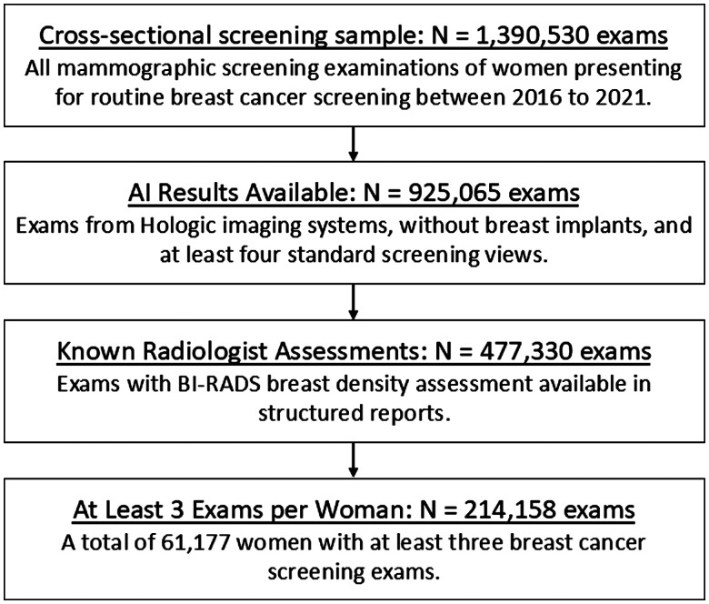
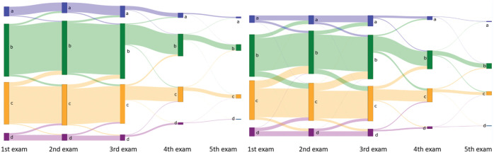
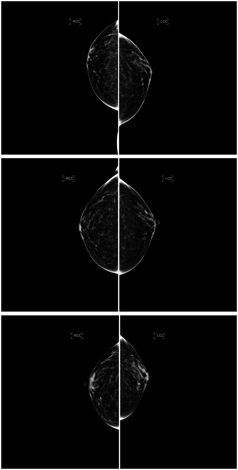
The AI model was evaluated retrospectively on a large mammography dataset including 50 sites across the United States from an outpatient radiology practice. Examinations were acquired on Hologic imaging systems between 2016 and 2021 and were interpreted by 39 radiologists (36% fellowship trained; years of experience: 2-37 years). Longitudinal patterns in 4-category breast density and binary breast density (non-dense vs. dense) were characterized for all women with at least 3 examinations (61 177 women; 214 158 examinations) as constant, descending, ascending, or bi-directional. Differences in longitudinal density patterns were assessed using paired proportion hypothesis testing.

RESULTS

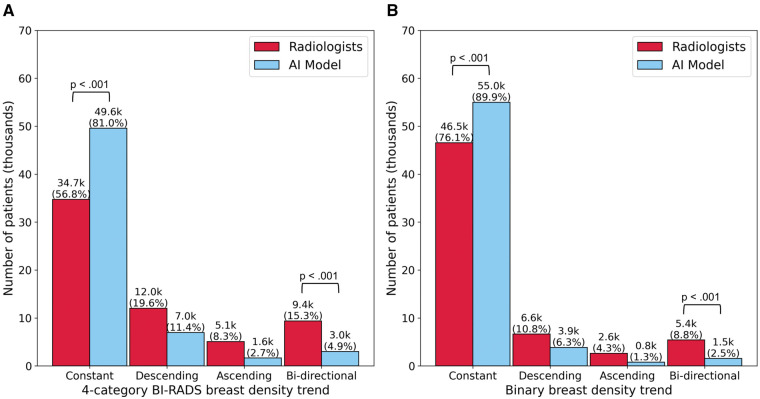
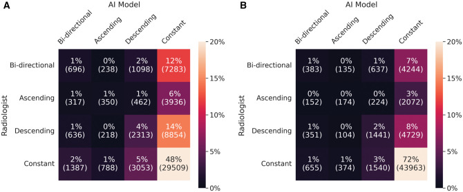
The AI model produced more constant ( < .001) and fewer bi-directional ( < .001) longitudinal density patterns compared to radiologists (AI: constant 81.0%, bi-directional 4.9%; radiologists: constant 56.8%, bi-directional 15.3%). The AI density model also produced more constant ( < .001) and fewer bi-directional ( < .001) longitudinal patterns for binary breast density. These findings held in various subset analyses, which minimize (1) change in breast density (post-menopausal women, women with stable image-based BMI), (2) inter-observer variability (same radiologist), and (3) variability by radiologist's training level (fellowship-trained radiologists).

CONCLUSIONS

AI produces more longitudinally consistent breast density assessments compared with interpreting radiologists.

ADVANCES IN KNOWLEDGE

Our results extend the advantages of AI in breast density evaluation beyond automation and reproducibility, showing a potential path to improved longitudinal consistency and more consistent downstream care for screened women.

摘要

目的

评估与放射科医生解读相比,使用人工智能(AI)模型进行乳房X光检查是否能带来更具纵向一致性的乳房密度评估。

方法

在一个大型乳房X光检查数据集上对AI模型进行回顾性评估,该数据集来自美国各地50个站点的门诊放射科实践。检查于2016年至2021年期间在Hologic成像系统上进行,由39名放射科医生进行解读(36%接受过专科培训;经验年限:2 - 37年)。对所有至少进行过3次检查的女性(61177名女性;214158次检查)的4类乳房密度和二元乳房密度(非致密型与致密型)的纵向模式进行特征描述,分为恒定、下降、上升或双向。使用配对比例假设检验评估纵向密度模式的差异。

结果

与放射科医生相比,AI模型产生的纵向密度模式更恒定(<0.001)且双向模式更少(<0.001)(AI:恒定81.0%,双向4.9%;放射科医生:恒定56.8%,双向15.3%)。AI密度模型在二元乳房密度方面也产生了更恒定(<0.001)且双向模式更少(<0.001)的纵向模式。这些发现在各种亚组分析中均成立,这些分析最小化了(1)乳房密度变化(绝经后女性、基于图像的BMI稳定的女性),(2)观察者间变异性(同一名放射科医生),以及(3)放射科医生培训水平的变异性(接受过专科培训的放射科医生)。

结论

与放射科医生解读相比,AI能产生更具纵向一致性的乳房密度评估。

知识进展

我们的结果将AI在乳房密度评估中的优势扩展到自动化和可重复性之外,显示出一条改善纵向一致性以及为筛查女性提供更一致的下游护理的潜在途径。

https://cdn.ncbi.nlm.nih.gov/pmc/blobs/34ee/11974406/1e5a3b50ac96/ubaf004f6.jpg
https://cdn.ncbi.nlm.nih.gov/pmc/blobs/34ee/11974406/516854e695f4/ubaf004f1.jpg
https://cdn.ncbi.nlm.nih.gov/pmc/blobs/34ee/11974406/56eaf5e25d12/ubaf004f2.jpg
https://cdn.ncbi.nlm.nih.gov/pmc/blobs/34ee/11974406/40f2f6759574/ubaf004f3.jpg
https://cdn.ncbi.nlm.nih.gov/pmc/blobs/34ee/11974406/68226d5fd05f/ubaf004f4.jpg
https://cdn.ncbi.nlm.nih.gov/pmc/blobs/34ee/11974406/42a143d698dd/ubaf004f5.jpg
https://cdn.ncbi.nlm.nih.gov/pmc/blobs/34ee/11974406/1e5a3b50ac96/ubaf004f6.jpg

相似文献

1
Artificial intelligence and consistency in patient care: a large-scale longitudinal study of mammographic density assessment.人工智能与患者护理的一致性:一项关于乳腺密度评估的大规模纵向研究
BJR Artif Intell. 2025 Mar 3;2(1):ubaf004. doi: 10.1093/bjrai/ubaf004. eCollection 2025 Jan.
2
Influence of AI Decision Support on Radiologists' Performance and Visual Search in Screening Mammography.人工智能决策支持对乳腺钼靶筛查中放射科医生表现及视觉搜索的影响
Radiology. 2025 Jul;316(1):e243688. doi: 10.1148/radiol.243688.
3
Mammographic density, endocrine therapy and breast cancer risk: a prognostic and predictive biomarker review.乳腺密度、内分泌治疗与乳腺癌风险:预后和预测生物标志物综述。
Cochrane Database Syst Rev. 2021 Oct 26;10(10):CD013091. doi: 10.1002/14651858.CD013091.pub2.
4
Mammography in combination with breast ultrasonography versus mammography for breast cancer screening in women at average risk.乳腺 X 线摄影联合乳腺超声与乳腺 X 线摄影用于一般风险女性乳腺癌筛查。
Cochrane Database Syst Rev. 2023 Mar 31;3(3):CD009632. doi: 10.1002/14651858.CD009632.pub3.
5
Variation within and between digital pathology and light microscopy for the diagnosis of histopathology slides: blinded crossover comparison study.数字病理学与光学显微镜检查在组织病理学切片诊断中的内部及相互间差异:双盲交叉对比研究
Health Technol Assess. 2025 Jul;29(30):1-75. doi: 10.3310/SPLK4325.
6
Sexual Harassment and Prevention Training性骚扰与预防培训
7
Artificial Intelligence and Radiologist Burnout.人工智能与放射科医生 burnout(职业倦怠)。
JAMA Netw Open. 2024 Nov 4;7(11):e2448714. doi: 10.1001/jamanetworkopen.2024.48714.
8
Cost-effectiveness of using prognostic information to select women with breast cancer for adjuvant systemic therapy.利用预后信息为乳腺癌患者选择辅助性全身治疗的成本效益
Health Technol Assess. 2006 Sep;10(34):iii-iv, ix-xi, 1-204. doi: 10.3310/hta10340.
9
External Validation of an Upgraded AI Model for Screening Ileocolic Intussusception Using Pediatric Abdominal Radiographs: Multicenter Retrospective Study.使用儿科腹部X光片筛查回结肠套叠的升级人工智能模型的外部验证:多中心回顾性研究
J Med Internet Res. 2025 Jul 8;27:e72097. doi: 10.2196/72097.
10
[Volume and health outcomes: evidence from systematic reviews and from evaluation of Italian hospital data].[容量与健康结果:来自系统评价和意大利医院数据评估的证据]
Epidemiol Prev. 2013 Mar-Jun;37(2-3 Suppl 2):1-100.

本文引用的文献

1
Patient Characteristics Impact False Positives in AI Interpretation of True-Negative Screening Breast Tomosynthesis Examinations.患者特征对乳腺断层合成阴性筛查检查人工智能解读中的假阳性结果产生影响。
Radiol Imaging Cancer. 2024 Sep;6(5):e249015. doi: 10.1148/rycan.249015.
2
Patient Characteristics Impact Performance of AI Algorithm in Interpreting Negative Screening Digital Breast Tomosynthesis Studies.患者特征影响 AI 算法解读阴性筛查数字乳腺断层摄影研究的性能。
Radiology. 2024 May;311(2):e232286. doi: 10.1148/radiol.232286.
3
Breast Density Evaluation According to BI-RADS 5th Edition on Digital Breast Tomosynthesis: AI Automated Assessment Versus Human Visual Assessment.
基于数字乳腺断层合成的BI-RADS第5版的乳腺密度评估:人工智能自动评估与人工视觉评估
J Pers Med. 2023 Mar 30;13(4):609. doi: 10.3390/jpm13040609.
4
Mammographic Screening in Routine Practice: Multisite Study of Digital Breast Tomosynthesis and Digital Mammography Screenings.常规实践中的乳腺 X 光筛查:数字乳腺断层合成术和数字乳腺 X 光筛查的多地点研究。
Radiology. 2023 May;307(3):e221571. doi: 10.1148/radiol.221571. Epub 2023 Mar 14.
5
Diagnostic accuracy of automated ACR BI-RADS breast density classification using deep convolutional neural networks.使用深度卷积神经网络的自动 ACR BI-RADS 乳腺密度分类的诊断准确性。
Eur Radiol. 2023 Jul;33(7):4589-4596. doi: 10.1007/s00330-023-09474-7. Epub 2023 Mar 1.
6
External Validation of an Ensemble Model for Automated Mammography Interpretation by Artificial Intelligence.人工智能辅助自动化乳腺 X 线摄影判读的集成模型的外部验证。
JAMA Netw Open. 2022 Nov 1;5(11):e2242343. doi: 10.1001/jamanetworkopen.2022.42343.
7
Association of Longitudinal Mammographic Breast Density Changes with Subsequent Breast Cancer Risk.纵向乳腺密度变化与随后乳腺癌风险的关联。
Radiology. 2023 Feb;306(2):e220291. doi: 10.1148/radiol.220291. Epub 2022 Sep 20.
8
Temporal quality degradation in AI models.人工智能模型中的时间质量下降。
Sci Rep. 2022 Jul 8;12(1):11654. doi: 10.1038/s41598-022-15245-z.
9
Artificial intelligence in mammographic phenotyping of breast cancer risk: a narrative review.人工智能在乳腺癌风险的乳腺摄影表型中的应用:叙述性综述。
Breast Cancer Res. 2022 Feb 20;24(1):14. doi: 10.1186/s13058-022-01509-z.
10
The impact of using weight estimated from mammographic images self-reported weight on breast cancer risk calculation.使用从乳腺钼靶图像估计的体重与自我报告的体重对乳腺癌风险计算的影响。
Proc SPIE Int Soc Opt Eng. 2017 Mar 3;10134. doi: 10.1117/12.2255619.