Lee Hsin-Ju, Lin Fa-Hsuan
Sunnybrook Research Institute, Toronto, Ontario, Canada.
Department of Medical Biophysics, University of Toronto, Toronto, Ontario, Canada.
Hum Brain Mapp. 2025 Apr 1;46(5):e70167. doi: 10.1002/hbm.70167.
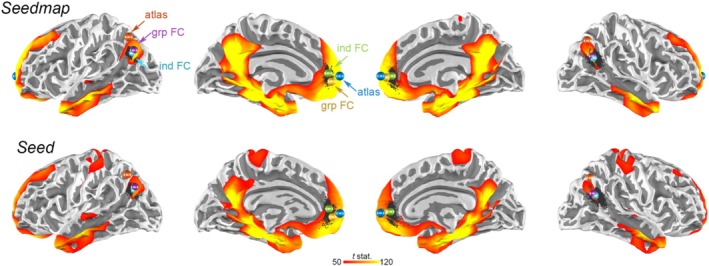
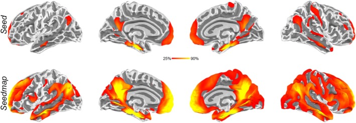
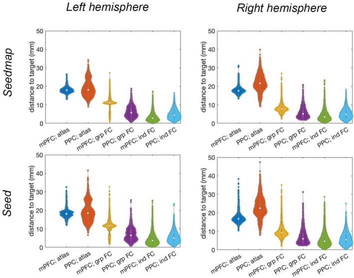
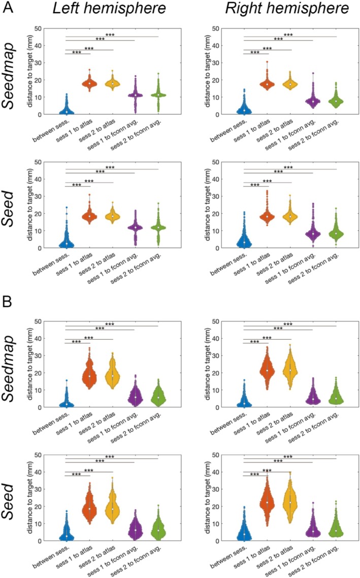
Individualized transcranial magnetic stimulation (TMS) targeting using functional connectivity analysis of functional magnetic resonance imaging (fMRI) has been demonstrated to be advantageous in inducing neuroplasticity. However, how this approach can benefit modulating the episodic memory function supported by the hippocampal network remains elusive. We use the resting-state fMRI data from a large cohort to reveal tentative TMS targets at cortical regions within the hippocampal network. Functional MRI from 1,133 individuals in the Human Connectome Project was used to analyze the hippocampal network using seed-based functional connectivity. Using a weighted sum of time series at the cortex, we identified the average centroids of individualized targets at the medial prefrontal cortex (mPFC) and posterior parietal cortices (PPCs) at (-10, 49, 7) and (-40, -67, 30) in the left hemisphere, respectively. The mPFC and PPC coordinate at the right hemispheres are (11, 51, 6) and (48, -59, 24) in the right hemisphere, respectively. Centroids of the individualized functional connectivity at the mPFC and PPC were reproducible between sessions with separations in average about 2 and 4 mm, respectively. These separations were significantly smaller than the distance to average functional connectivity centroids (10 mm) and atlas coordinate (20 mm). These coordinates can be reliably identified (> 90% of individuals) using cortical "seedmaps." Our results suggest candidate TMS target coordinates to modulate the hippocampal function.
使用功能磁共振成像(fMRI)的功能连接分析进行个体化经颅磁刺激(TMS)靶向已被证明在诱导神经可塑性方面具有优势。然而,这种方法如何有助于调节海马网络支持的情景记忆功能仍不清楚。我们使用来自一大群人的静息态fMRI数据来揭示海马网络内皮质区域的初步TMS靶点。人类连接组计划中1133名个体的功能磁共振成像用于通过基于种子的功能连接分析海马网络。使用皮质处时间序列的加权和,我们分别在左半球的内侧前额叶皮质(mPFC)和顶叶后皮质(PPCs)中确定了个体化靶点的平均质心,坐标分别为(-10, 49, 7)和(-40, -67, 30)。右半球的mPFC和PPC坐标分别为(11, 51, 6)和(48, -59, 24)。mPFC和PPC处个体化功能连接的质心在各次会话之间具有可重复性,平均间隔分别约为2和4毫米。这些间隔明显小于到平均功能连接质心(约10毫米)和图谱坐标(约20毫米)的距离。使用皮质“种子图”可以可靠地识别这些坐标(超过90%的个体)。我们的结果表明了调节海马功能的候选TMS靶点坐标。