Zheng Zihui, Nie Aobo, Wu Xiaojie, Chen Shi, Zhang Lijun, Yang Dongqing, Shi Yuqing, Xiong Xiyu, Guo Jun
Department of Biochemistry and Molecular Biology, School of Medicine, Nanjing University of Chinese Medicine, Nanjing, Jiangsu, 210023, People's Republic of China.
State Key Laboratory on Technologies for Chinese Medicine Pharmaceutical Process Control and Intelligent Manufacture, Nanjing University of Chinese Medicine, Nanjing, 210023, People's Republic of China.
Int J Nanomedicine. 2025 Apr 5;20:4145-4163. doi: 10.2147/IJN.S503181. eCollection 2025.
Osmotic imbalance is a critical driving force of cerebral edema. Protein nanoparticles (PNs) amplify intracellular osmotic effects by regulating membrane potential and homeostasis of water and multiple ions. This study has investigated how PNs control the neuronal swelling through electromechanical activity.
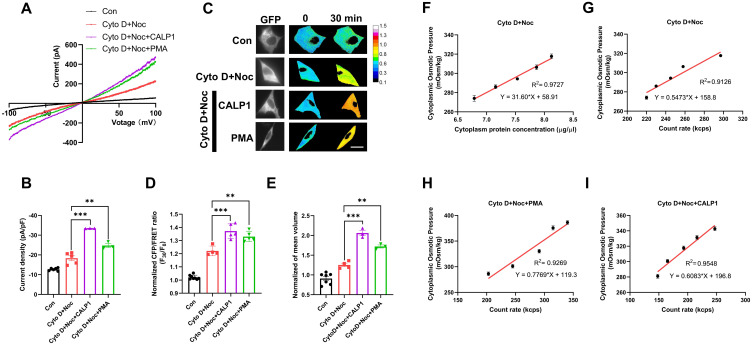
The fluorescence resonance energy transfer (FRET)-based Vimentin force probe was used to real-time monitor the osmotic tension in neurons. Patch clamp and the living cell 3D imaging system were applied to explore the relationship between cell electromechanical activity and cell volume in different cytotoxic cell models. Cytoplasmic PN amount measured by the NanoSight instrument, ion contents detected by the freezing point osmometer and ion imaging were performed to investigate the role of PNs in regulating cell swelling.
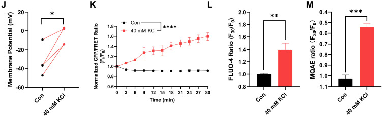
We observed a close association between neuronal swelling and changes in osmotic tension and membrane potential. The tension effect of biological osmotic pressure (OP) relies on electromechanical cooperation induced by intracellular PN and Ca levels. PNs increment results from cytoplasmic translocation of intracellular various proteins. Alterations in Ca content are involved in the membrane potential transition between depolarization and hyperpolarization in a PN-dependent manner. Chemical signals-mediated sensitization of ion channels has an indispensable effect on PN-induced ion increments. Notably, aquaporin-mediated water influx recovers membrane potential and enhances osmotic tension controlling neuronal swelling.
Our findings indicate that PNs, Ca, and water are pivotal in electromechanical cooperation and provide insights into the biological OP mechanisms underlying neurotoxic edema.
渗透失衡是脑水肿的关键驱动力。蛋白质纳米颗粒(PNs)通过调节膜电位以及水和多种离子的稳态来放大细胞内的渗透效应。本研究探讨了PNs如何通过机电活性控制神经元肿胀。
基于荧光共振能量转移(FRET)的波形蛋白力探针用于实时监测神经元中的渗透压。膜片钳和活细胞3D成像系统用于探究不同细胞毒性细胞模型中细胞机电活性与细胞体积之间的关系。采用纳米可视仪器测量细胞质PN含量,用冰点渗透压计检测离子含量并进行离子成像,以研究PNs在调节细胞肿胀中的作用。
我们观察到神经元肿胀与渗透压和膜电位变化之间存在密切关联。生物渗透压(OP)的张力效应依赖于细胞内PN和钙水平诱导的机电协同作用。PNs的增加源于细胞内各种蛋白质的细胞质转位。钙含量的变化以PN依赖的方式参与去极化和超极化之间的膜电位转变。化学信号介导的离子通道敏化对PN诱导的离子增加具有不可或缺的作用。值得注意的是,水通道蛋白介导的水流入恢复膜电位并增强控制神经元肿胀的渗透压。
我们的研究结果表明,PNs、钙和水在机电协同作用中起关键作用,并为神经毒性水肿潜在的生物OP机制提供了见解。