Denis Louise, Meseguer Elena, Gaudemer Augustin, Jaklh Georges, Bodard Sylvain, Chabouh Georges, Hervé Dominique, Vicaut Eric, Amarenco Pierre, Couture Olivier
Sorbonne Université, CNRS, INSERM Laboratoire d'Imagerie Biomédicale, Paris, France.
Department of Neurology Bichat University Hospital (APHP), Paris, France.
Theranostics. 2025 Mar 10;15(9):4074-4083. doi: 10.7150/thno.105427. eCollection 2025.
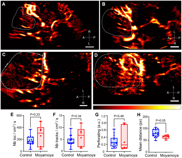
Deep brain structures are supplied by perforating arteries, which are too thin to be observed with non-invasive and widely available clinical imaging methods. In moyamoya disease, main arteries in the base of the brain progressively narrow, and perforating arteries grow densely and tortuously to compensate the lack of blood supply in deep brain structures. The aim of this study is to evaluate the efficacy of transcranial ultrasound localization microscopy (ULM) in visualizing perforating arteries, utilizing a standard low-frame-rate ultrasound clinical scanner and contrast sequences commonly employed in hospital settings. This prospective single-center study included ischemic stroke patients not related to the study of perforating arteries, and moyamoya disease patients. Contrast-enhanced ultrasound sequences (CEUS) were performed by an experienced neurologist and the images acquired were used to perform post-processing ULM. ULM density maps were compared with conventional 3T TOF MRI and color Doppler imaging in both groups. We included a group of 15 control patients and another group of 9 moyamoya patients between March 2023 and March 2024. The patients had an average age of 45 ± 14 years (65% male). Perforating arteries were captured on all subjects, with a mean diameter of 0.8 ± 0.3 mm in control patients, while it was not possible with TOF MRI or color Doppler (P < 0.05). Moreover, ULM enabled to highlight differences between healthy subjects and those with moyamoya disease through track mean distance (P = 0.05). Using a low-frame-rate ultrasound scanner, CEUS and accessible post-processing tools, we demonstrate that transcranial ULM can facilitate the visualization and characterization of perforating arteries, even in cases where they were previously undetectable using standard non-invasive imaging techniques. We speculate that with the advent of high-frame-rate 3D ULM, this technique may find widespread utility in hospitals.
深部脑结构由穿支动脉供血,这些动脉过于纤细,无法用非侵入性且广泛应用的临床成像方法观察到。在烟雾病中,脑底部的主要动脉逐渐变窄,穿支动脉则密集且迂曲地生长,以补偿深部脑结构血供的不足。本研究的目的是利用标准的低帧率超声临床扫描仪和医院常用的造影序列,评估经颅超声定位显微镜(ULM)在显示穿支动脉方面的有效性。这项前瞻性单中心研究纳入了与穿支动脉研究无关的缺血性中风患者和烟雾病患者。由经验丰富的神经科医生进行对比增强超声序列(CEUS)检查,并将采集到的图像用于进行后处理ULM。在两组中,将ULM密度图与传统的3T TOF MRI和彩色多普勒成像进行比较。在2023年3月至2024年3月期间,我们纳入了一组15名对照患者和另一组9名烟雾病患者。患者的平均年龄为45±14岁(65%为男性)。在所有受试者中均捕捉到了穿支动脉,对照患者的平均直径为0.8±0.3毫米,而TOF MRI或彩色多普勒则无法做到(P<0.05)。此外,ULM能够通过轨迹平均距离突出健康受试者与烟雾病患者之间的差异(P = 0.05)。使用低帧率超声扫描仪、CEUS和可获取的后处理工具,我们证明经颅ULM能够促进穿支动脉的可视化和特征描述,即使在使用标准非侵入性成像技术之前无法检测到它们的情况下也是如此。我们推测,随着高帧率3D ULM的出现,这项技术可能会在医院中得到广泛应用。