Torun Raziye, Golbasi Hakan, Saglam Ceren, Tuncer Can Sevim, Gercik Ilayda, Ankara Aktas Hale, Toka Ilknur, Emiralioglu Cakir Zubeyde, Sengul Mustafa, Ekin Atalay
Department of Perinatology, Izmir City Hospital, 35510 Izmir, Turkey.
Department of Obstetrics and Gynecology, Faculty of Medicine, Izmir Katip Celebi University, 35620 Izmir, Turkey.
J Clin Med. 2025 Mar 24;14(7):2204. doi: 10.3390/jcm14072204.
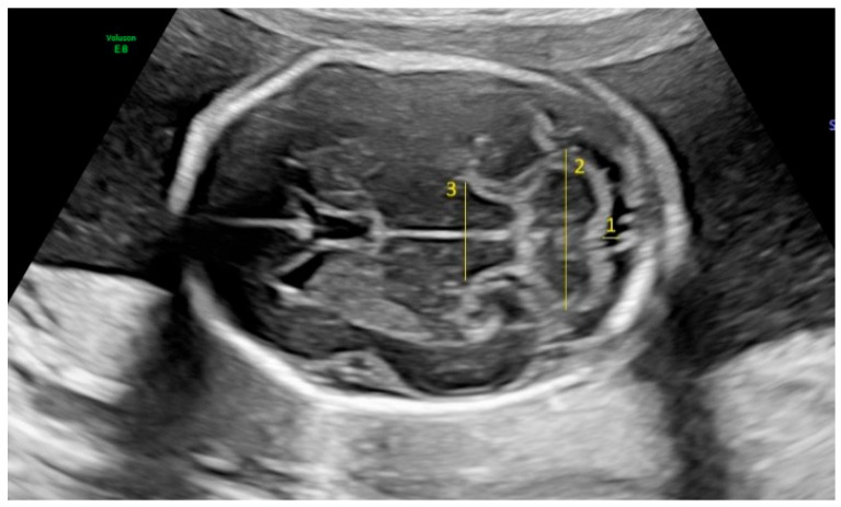
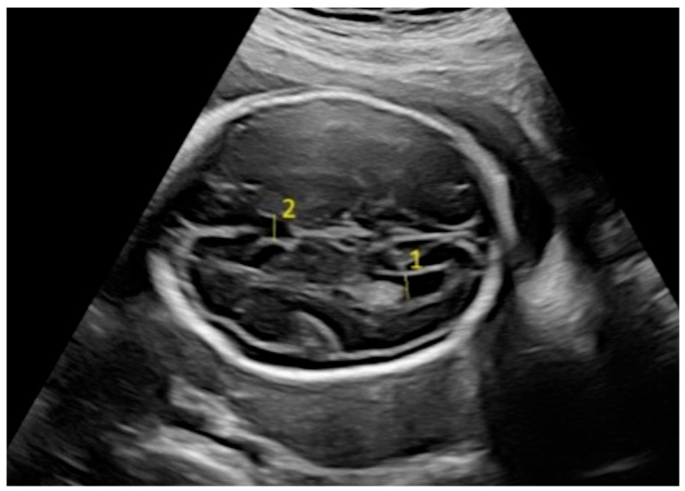
: Maternal thyroid function plays a crucial role in fetal brain development, yet the potential impact of maternal hypothyroidism and thyroid autoimmunity on fetal intracranial structures remains inadequately explored. To investigate the impact of maternal hypothyroidism and thyroid autoimmunity on fetal intracranial structures, focusing on potential alterations in critical brain parameters during mid-gestation. : This prospective case-control study included pregnant women between 18 and 24 weeks of gestation. Participants were divided into three groups: hypothyroidism and antibodies (Ab) group, hypothyroidism and Ab(-) group, and the control group. Ultrasonographic measurements of fetal intracranial structures such as the posterior lateral ventricle (PLV), cavum septum pellucidi (CSP), cisterna magna (CM), thalamus, and transcerebellar diameter (TCD) were recorded and compared. : A total of 153 pregnant women were evaluated (n = 52 in the hypothyroidism and Ab(+) group, n = 51 in the hypothyroidism and Ab(-) group, and n = 50 in the control group). Although most of the biometric parameters were similar across the groups, the hypothyroidism and Ab(+) group exhibited significantly lower PLV and thalamus measurements compared to the control group ( < 0.05). Additionally, there was a notable difference in the BMI among the groups, with hypothyroid participants (with or without antibodies) showing higher rates of being overweight or obese. : Maternal hypothyroidism and the presence of thyroid autoantibodies may be associated with subtle changes in fetal brain structures during the mid-gestation period, particularly in the thalamus and PLV.
母体甲状腺功能在胎儿脑发育中起着至关重要的作用,然而,母体甲状腺功能减退和甲状腺自身免疫对胎儿颅内结构的潜在影响仍未得到充分研究。为了研究母体甲状腺功能减退和甲状腺自身免疫对胎儿颅内结构的影响,重点关注妊娠中期关键脑参数的潜在变化。:这项前瞻性病例对照研究纳入了妊娠18至24周的孕妇。参与者分为三组:甲状腺功能减退和抗体(Ab)组、甲状腺功能减退和Ab(-)组以及对照组。记录并比较胎儿颅内结构如侧脑室后角(PLV)、透明隔腔(CSP)、枕大池(CM)、丘脑和小脑横径(TCD)的超声测量值。:总共评估了153名孕妇(甲状腺功能减退和Ab(+)组n = 52,甲状腺功能减退和Ab(-)组n = 51,对照组n = 50)。尽管大多数生物测量参数在各组之间相似,但与对照组相比,甲状腺功能减退和Ab(+)组的PLV和丘脑测量值显著更低(<0.05)。此外,各组之间的BMI存在显著差异,甲状腺功能减退的参与者(有或没有抗体)超重或肥胖的发生率更高。:母体甲状腺功能减退和甲状腺自身抗体的存在可能与妊娠中期胎儿脑结构的细微变化有关,特别是在丘脑和PLV。