Wagle Laxman, Timshina Anuj, Pant Hom N, Pathak Vikas
Internal Medicine, Ascension Saint Agnes Hospital, Baltimore, USA.
Internal Medicine, MedStar Franklin Square Medical Center, Baltimore, USA.
Cureus. 2025 Mar 13;17(3):e80528. doi: 10.7759/cureus.80528. eCollection 2025 Mar.
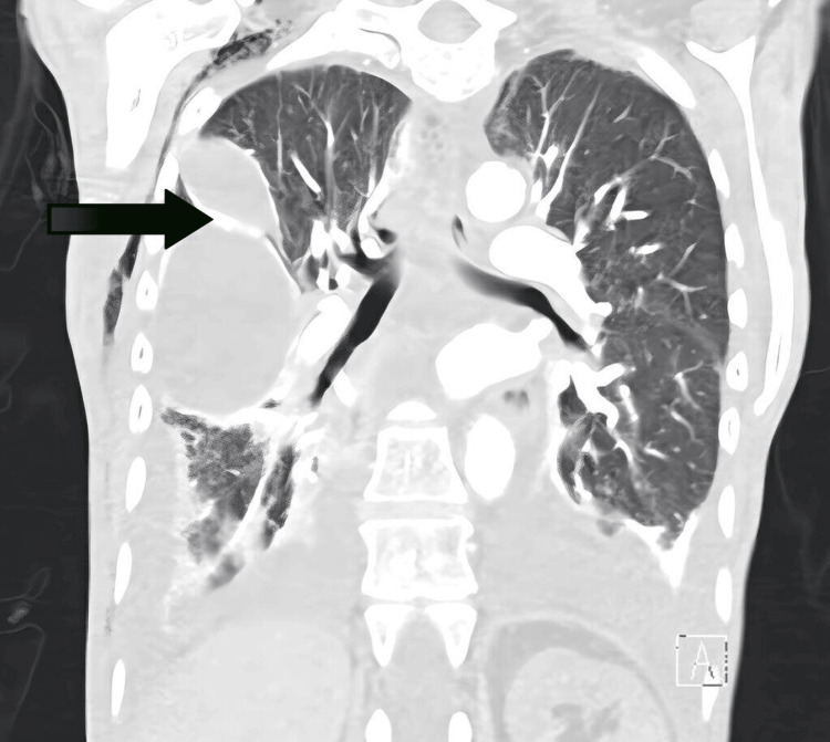
Radiation therapy (RT) is a common treatment for non-Hodgkin lymphoma (NHL) but can lead to long-term pulmonary and cardiovascular complications. Delayed radiotherapy-related pleural effusion (DRPE) and pericardial effusion are rare sequelae, with few cases reported. This case highlights recurrent pleural and pericardial effusions nearly 40 years after chest RT, underscoring the need for ongoing surveillance in cancer survivors. A 51-year-old female with a history of nodular sclerosing NHL in remission after RT in 1977 presented in 2016 with recurrent bilateral pleural and pericardial effusions. Despite multiple interventions, including pericardiocentesis, thoracenteses, and pleural catheter placement, her effusions persisted. An extensive workup ruled out malignancy, infection, and autoimmune causes, ultimately attributing the effusions to radiation-induced lung injury. Despite ongoing management, she was discharged to hospice care. DRPE is a diagnostic challenge due to its delayed onset, sometimes appearing decades after RT. It can present with variable pleural fluid characteristics. Radiation-induced lung injury is a known complication of thoracic RT, with risk factors including radiation dose and pre-existing pulmonary conditions. Management is symptomatic, with treatments such as NSAIDs, corticosteroids, diuretics, and pleural drainage, though outcomes vary. In this case, pleural catheter placement offered temporary relief, but recurrent effusions led to hospice care. This case highlights the need for long-term monitoring in survivors of chest RT, as delayed pulmonary and cardiovascular toxicities can arise decades later. Given the rarity of DRPE and the lack of standardized treatment, further research into protective strategies and early interventions for radiation-induced lung injury is essential to improve cancer survivors' quality of life.
放射治疗(RT)是非霍奇金淋巴瘤(NHL)的常见治疗方法,但可导致长期的肺部和心血管并发症。放疗相关的迟发性胸腔积液(DRPE)和心包积液是罕见的后遗症,报道的病例很少。本病例强调了胸部放疗近40年后复发性胸腔和心包积液,突出了癌症幸存者持续监测的必要性。一名51岁女性,有结节硬化型NHL病史,1977年放疗后缓解,2016年出现复发性双侧胸腔和心包积液。尽管进行了多次干预,包括心包穿刺、胸腔穿刺和胸腔置管,她的积液仍持续存在。广泛检查排除了恶性肿瘤、感染和自身免疫性病因,最终将积液归因于放射性肺损伤。尽管持续治疗,她还是出院接受临终关怀。DRPE因其发病延迟,有时在放疗后数十年出现,是一个诊断挑战。它可表现为胸腔积液特征各异。放射性肺损伤是胸部放疗已知的并发症,危险因素包括辐射剂量和既往肺部疾病。治疗以对症为主,如使用非甾体抗炎药、皮质类固醇、利尿剂和胸腔引流等,不过疗效各异。在本病例中,胸腔置管提供了暂时缓解,但复发性积液导致了临终关怀。本病例强调了胸部放疗幸存者长期监测的必要性,因为迟发性肺部和心血管毒性可能在数十年后出现。鉴于DRPE的罕见性和缺乏标准化治疗,进一步研究放射性肺损伤的保护策略和早期干预对于提高癌症幸存者的生活质量至关重要。