Hussein Sara M, Rames Jess D, Shehab Abdallah A, Pazelli Alexandre M, Sears Victoria A, Wentworth Adam J, Morris Jonathan M, Sharaf Basel A
From the Division of Plastic and Reconstructive Surgery, Department of Surgery, Mayo Clinic, Rochester, MN.
Neural Engineering and Precision Surgery Laboratorie, Mayo Clinic, Rochester, MN.
Plast Reconstr Surg Glob Open. 2025 Apr 21;13(4):e6650. doi: 10.1097/GOX.0000000000006650. eCollection 2025 Apr.
Mandible contour significantly influences facial appearance, framing the lower facial silhouette. Redefining mandibular contour is key for facial and neck rejuvenation. Yet, there is limited facial aging research across different lifespans and sexes. Here, we utilize artificial intelligence and advanced 3-dimensional (3D) analysis to elucidate mandibular aging patterns in male and female subjects.
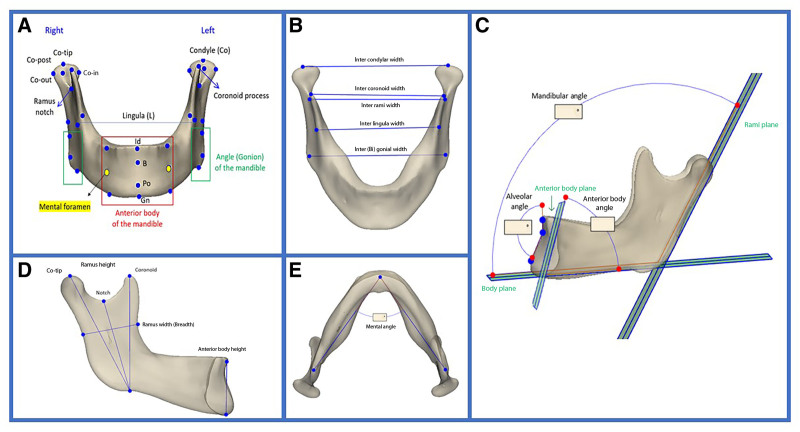
A retrospective analysis of facial computed tomography scans in White patients was conducted, categorizing subjects into 3 age groups (20-79 y) and stratifying them by sex. Artificial intelligence-assisted segmentation into 3D mandibles was done in Mimics v.25, and statistical shape modeling was used to create an average mandible for each group. Volume and linear measurements were assessed via 3D overlays.
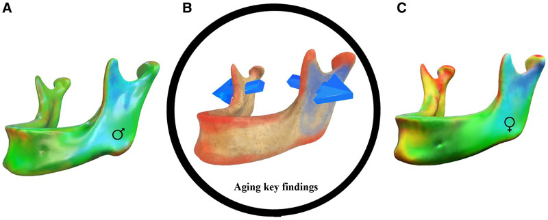
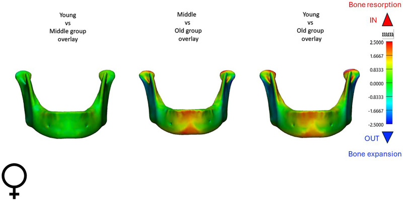
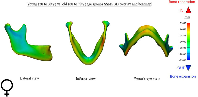
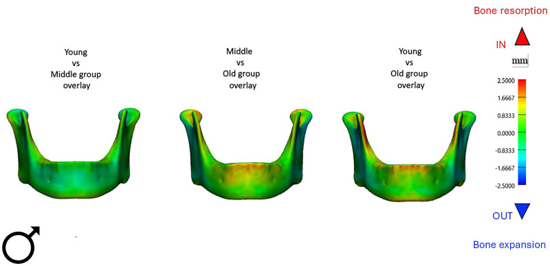
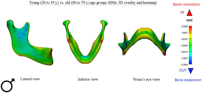
Analysis of 280 mandibles demonstrated statistically significant aging changes in both sexes. Ramus height showed a marked decrease with age, by approximately 5.3 mm in women and 4.2 mm in men ( < 0.001). Interrami and intercondylar widths increased by a mean of 4-5 mm ( < 0.01). Women exhibited an increase in mandibular angle ( < 0.01), and bony resorption over the chin compared to men, who exhibited concentrated bone resorption at the gonion projection.
Mandibular aging, independent of tooth loss, exhibits specific bone remodeling patterns by sex. Posteriorly, mandibular widths increase in both sexes, whereas ramus height decreases. Women experience more resorption at the anterior alveolar surface and chin than men. Statistical shape modeling effectively visualizes these patterns on a population level, bridging the gap between traditional aging research and current understanding.
下颌轮廓显著影响面部外观,勾勒出面部下部轮廓。重新塑造下颌轮廓是面部和颈部年轻化的关键。然而,针对不同寿命阶段和性别的面部衰老研究有限。在此,我们利用人工智能和先进的三维(3D)分析来阐明男性和女性受试者的下颌衰老模式。
对白人患者的面部计算机断层扫描进行回顾性分析,将受试者分为3个年龄组(20 - 79岁)并按性别分层。在Mimics v.25软件中通过人工智能辅助将其分割为3D下颌骨,并使用统计形状建模为每个组创建平均下颌骨。通过3D叠加评估体积和线性测量值。
对280个下颌骨的分析表明,两性均存在具有统计学意义的衰老变化。下颌支高度随年龄显著降低,女性约降低5.3毫米,男性约降低4.2毫米(<0.001)。下颌支间宽度和髁突间宽度平均增加4 - 5毫米(<0.01)。女性的下颌角增大(<0.01),与男性相比,女性下巴处有骨质吸收,而男性在角突处有集中的骨质吸收。
下颌衰老与牙齿缺失无关,按性别表现出特定的骨重塑模式。在后部,两性的下颌宽度均增加,而下颌支高度降低。女性在前牙槽表面和下巴处的吸收比男性更多。统计形状建模有效地在群体水平上可视化了这些模式,弥合了传统衰老研究与当前认识之间的差距。