Liao Bin, Wang Pan, Gong Sheng, Zhao Lu, Liu Jie, Wu Nan
Chongqing Medical University, Chongqing, China.
Department of Neurosurgery, Chongqing Research Center for Glioma Precision Medicine, Chongqing General Hospital, Chongqing University, Chongqing, China.
Front Pharmacol. 2025 Apr 11;16:1575332. doi: 10.3389/fphar.2025.1575332. eCollection 2025.
This study investigates whether Hypoxia-Inducible Factor 1 alpha (HIF1α) and Hypoxia-Inducible Factor 2 alpha (HIF2α) coordinately regulate insulin-like growth factor 1 receptor (IGF1R) expression, thereby influencing chemosensitivity in glioblastoma multiforme (GBM).
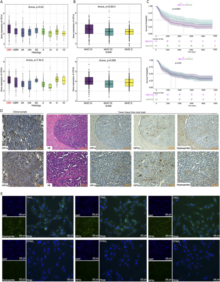
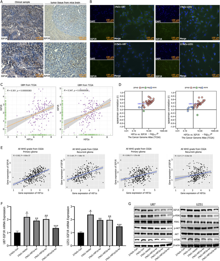
We analyzed the expression and correlation of HIF1α, HIF2α, and IGF1R in glioma using The Cancer Genome Atlas (TCGA) and Chinese Glioma Genome Atlas (CGGA) databases. Immunohistochemistry (IHC) was performed to detect the expression of HIF1α, HIF2α, and IGF1R in GBM tissues and cells, as well as oxygen tension. Cell cycle analysis, apoptosis assays, lactate dehydrogenase (LDH) release measurements, Western blotting, and xenograft tumor models were employed to explore the synergistic regulation of IGF1R by HIF1α and HIF2α, focusing on activation of the PI3K/AKT signaling pathway and its contribution to GBM drug resistance. Chromatin immunoprecipitation-quantitative PCR (ChIP-qPCR) and dual-luciferase reporter assays were used to investigate the binding sites of HIF1α and HIF2α involved in regulating IGF1R.
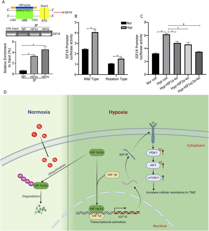
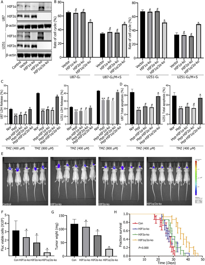
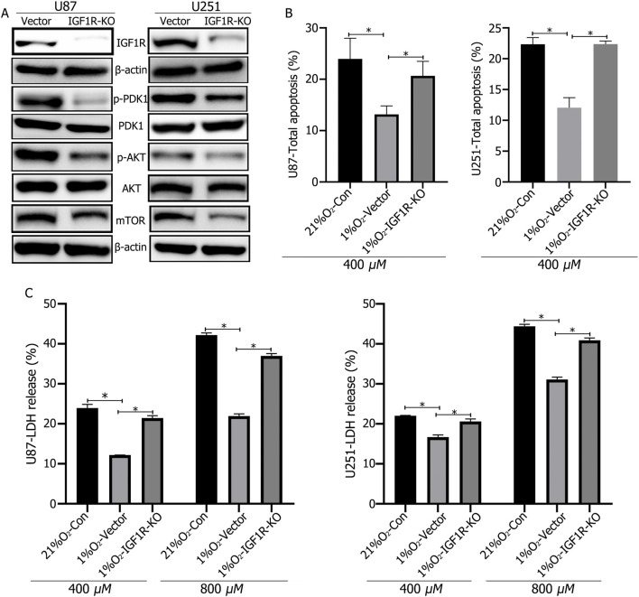
Our study demonstrated that HIF1α and HIF2α were highly expressed in GBM tissues and hypoxia-cultured cells, and their expression positively correlated with IGF1R expression. Simultaneous knockout of HIF1α and HIF2α in GBM cells resulted in the highest LDH release and apoptosis rates under hypoxic conditions, accompanied by the most significant decrease in IGF1R, p-PDK1, and p-AKT levels. Knockout of IGF1R in tumor cells under hypoxia led to an increas of LDH release and apoptosis rates, and reduced phosphorylation of PDK1 and AKT. In addition, we demonstrated that HIF1α and HIF2α promoted IGF1R expression by binding to a specific hypoxia response element (HRE) sequence (5'-GAACGTGCCT-3') within the IGF1R promoter using dual-luciferase reporter system.
Glioblastoma cells, residing within a hypoxic microenvironment, exhibit high expression of HIF1α and HIF2α. These transcription factors promote the upregulation of IGF1R, which subsequently activates the PI3K/AKT signaling pathway. This activation, in turn, promotes cell proliferation and chemoresistance, ultimately contributing to tumor malignancy.
本研究调查缺氧诱导因子1α(HIF1α)和缺氧诱导因子2α(HIF2α)是否协同调节胰岛素样生长因子1受体(IGF1R)的表达,从而影响多形性胶质母细胞瘤(GBM)的化疗敏感性。
我们使用癌症基因组图谱(TCGA)和中国胶质瘤基因组图谱(CGGA)数据库分析了胶质瘤中HIF1α、HIF2α和IGF1R的表达及相关性。进行免疫组织化学(IHC)检测GBM组织和细胞中HIF1α、HIF2α和IGF1R的表达以及氧张力。采用细胞周期分析、凋亡检测、乳酸脱氢酶(LDH)释放测定、蛋白质免疫印迹法和异种移植肿瘤模型,探讨HIF1α和HIF2α对IGF1R的协同调节作用,重点关注PI3K/AKT信号通路的激活及其对GBM耐药性的影响。采用染色质免疫沉淀-定量聚合酶链反应(ChIP-qPCR)和双荧光素酶报告基因检测法研究HIF1α和HIF2α参与调节IGF1R的结合位点。
我们的研究表明,HIF1α和HIF2α在GBM组织和缺氧培养的细胞中高表达,它们的表达与IGF1R表达呈正相关。在GBM细胞中同时敲除HIF1α和HIF2α导致缺氧条件下LDH释放率和凋亡率最高,同时IGF1R、p-PDK1和p-AKT水平显著降低。缺氧条件下肿瘤细胞中IGF1R的敲除导致LDH释放率和凋亡率增加,PDK1和AKT的磷酸化减少。此外,我们使用双荧光素酶报告系统证明,HIF1α和HIF2α通过与IGF1R启动子内的特定缺氧反应元件(HRE)序列(5'-GAACGTGCCT-3')结合来促进IGF1R的表达。
存在于缺氧微环境中的胶质母细胞瘤细胞表现出HIF1α和HIF2α的高表达。这些转录因子促进IGF1R的上调,随后激活PI3K/AKT信号通路。这种激活反过来促进细胞增殖和化疗耐药性,最终导致肿瘤恶性进展。