Palka Océane, Guillin Raphaël, Lecigne Romain, Combes Damien
Department of Radiology, University Hospital of Rennes, Rennes, France.
Department of Radiology, University Hospital of Angers, Angers, France.
Insights Imaging. 2025 Apr 29;16(1):94. doi: 10.1186/s13244-025-01945-3.
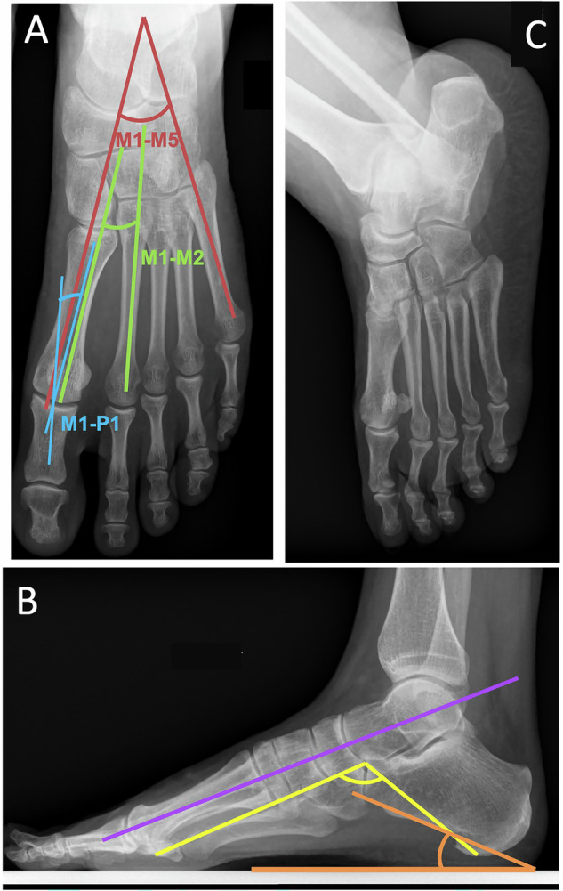
Metatarsalgia, characterized by forefoot pain, is frequent and is primarily due to foot static disorders. Initial evaluation with weight-bearing radiographs is essential, allowing precise analysis of the architecture of the foot. Ultrasound is useful for soft tissue and tendon examination and provides the best clinical correlation. Computed Tomography provides detailed bone assessment and is helpful for pre-operative planning. Magnetic Resonance Imaging is the gold standard modality, offering superior soft tissue contrast. The common causes of metatarsalgia include hallux pathologies (hallux valgus, hallux rigidus, and sesamoid issues), bursitis (intermetatarsal and subcapitellar), Morton's neuroma, second ray syndrome, stress fractures, and systemic pathologies affecting the foot. Combining clinical and imaging data is crucial for accurate diagnosis and effective management of metatarsalgia. Post-traumatic causes of metatarsalgia are beyond the scope of this article and will not be described. CRITICAL RELEVANCE STATEMENT: Metatarsalgia, the pain of the forefoot, necessitates accurate imaging for diagnosis and management. This review critically assesses imaging techniques and diagnostic approaches, aiming to enhance radiological practice and support effective therapeutic decision-making. KEY POINTS: Metatarsalgia commonly results from foot static disorders, requiring weight-bearing radiographs for assessment. MRI is often the gold standard examination, but ultrasound is complementary, allowing for a radioclinical approach with dynamic examinations. The radiologist is crucial in diagnosing metatarsalgia, providing essential imaging, and guiding treatment.
跖痛症以足前部疼痛为特征,较为常见,主要由足部静态紊乱引起。负重X线片的初步评估至关重要,有助于精确分析足部结构。超声对软组织和肌腱检查有用,并能提供最佳的临床相关性。计算机断层扫描可提供详细的骨骼评估,有助于术前规划。磁共振成像为金标准检查方式,能提供卓越的软组织对比度。跖痛症的常见病因包括拇趾病变(拇外翻、拇僵硬症和籽骨问题)、滑囊炎(跖间和跖骨头下)、莫顿神经瘤、第二跖骨综合征、应力性骨折以及影响足部的全身性病变。结合临床和影像学数据对于跖痛症的准确诊断和有效管理至关重要。创伤后跖痛症的病因不在本文讨论范围内,故不予描述。关键相关性声明:跖痛症,即足前部疼痛,诊断和管理需要准确的影像学检查。本综述批判性地评估了影像学技术和诊断方法,旨在提高放射学实践水平并支持有效的治疗决策。要点:跖痛症通常由足部静态紊乱导致,评估需要负重X线片。磁共振成像通常是金标准检查,但超声具有辅助作用,可通过动态检查实现放射临床方法。放射科医生在跖痛症的诊断、提供必要的影像学检查及指导治疗方面至关重要。