• 文献检索
  • 文档翻译
  • 深度研究
  • 学术资讯
  • Suppr Zotero 插件Zotero 插件
  • 邀请有礼
  • 套餐&价格
  • 历史记录
应用&插件
Suppr Zotero 插件Zotero 插件浏览器插件Mac 客户端Windows 客户端微信小程序
定价
高级版会员购买积分包购买API积分包
服务
文献检索文档翻译深度研究API 文档MCP 服务
关于我们
关于 Suppr公司介绍联系我们用户协议隐私条款
关注我们

Suppr 超能文献

核心技术专利:CN118964589B侵权必究
粤ICP备2023148730 号-1Suppr @ 2026

文献检索

告别复杂PubMed语法,用中文像聊天一样搜索,搜遍4000万医学文献。AI智能推荐,让科研检索更轻松。

立即免费搜索

文件翻译

保留排版,准确专业,支持PDF/Word/PPT等文件格式,支持 12+语言互译。

免费翻译文档

深度研究

AI帮你快速写综述,25分钟生成高质量综述,智能提取关键信息,辅助科研写作。

立即免费体验

前列腺微钙化晶体学作为病理学标志物

Prostate microcalcification crystallography as a marker of pathology.

作者信息

Gosling Sarah B, Arnold Emily L, Adams Lois, Cool Paul, Geraki Kalotina, Kitchen Mark O, Lyburn Iain D, Rogers Keith D, Snow Tim, Stone Nick, Greenwood Charlene E

机构信息

School of Chemical and Physical Sciences, Keele University, Keele, Staffordshire, ST5 5BG, UK.

Diamond Light Source, Harwell Science and Innovation Campus, Didcot, OX11 0DE, UK.

出版信息

Sci Rep. 2025 Apr 29;15(1):14979. doi: 10.1038/s41598-025-98692-8.

DOI:10.1038/s41598-025-98692-8
PMID:40301587
原文链接:https://pmc.ncbi.nlm.nih.gov/articles/PMC12041448/
Abstract

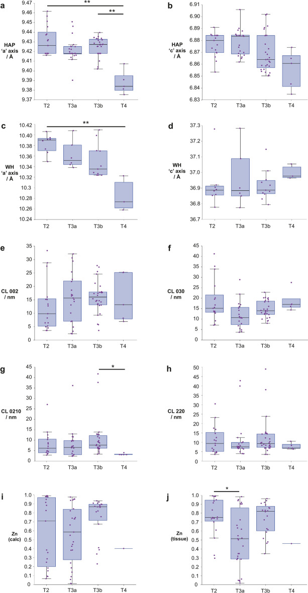
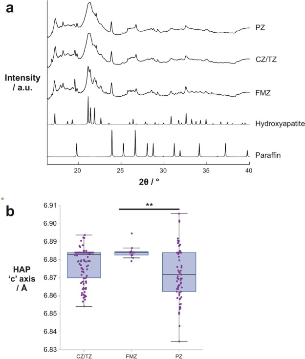
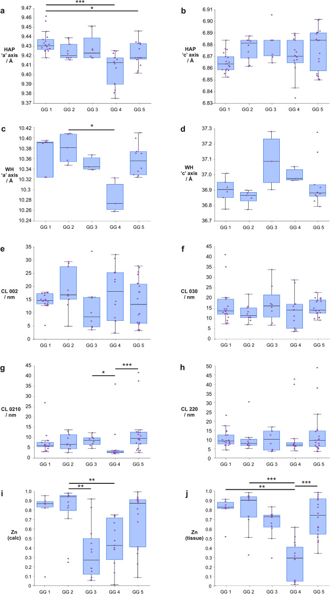
Prostate cancer remains the most common male cancer; however, treatment regimens remain unclear in some cases due to a lack of agreement in current testing methods. Therefore, there is an increasing need to identify novel biomarkers to better counsel patients about their treatment options. Microcalcifications offer one such avenue of exploration. Microfocus spectroscopy at the i18 beamline at Diamond Light Source was utilised to measure X-ray diffraction and fluorescence maps of calcifications in 10 µm thick formalin fixed paraffin embedded prostate sections. Calcifications predominantly consisted of hydroxyapatite (HAP) and whitlockite (WH). Kendall's Tau statistics showed weak correlations of 'a' and 'c' lattice parameters in HAP with GG (r = - 0.323, p = 3.43 × 10 and r = 0.227, p = 0.011 respectively), and a negative correlation of relative zinc levels in soft tissue (r = - 0.240, p = 0.022) with GG. Negative correlations of the HAP 'a' axis (r = - 0.284, p = 2.17 × 10) and WH 'c' axis (r = - 0.543, p = 2.83 × 10) with pathological stage were also demonstrated. Prostate calcification chemistry has been revealed for the first time to correlate with clinical markers, highlighting the potential of calcifications as biomarkers of prostate cancer.

摘要

前列腺癌仍然是最常见的男性癌症;然而,由于目前检测方法存在分歧,某些情况下的治疗方案仍不明确。因此,越来越需要识别新的生物标志物,以便更好地为患者提供治疗选择方面的建议。微钙化提供了这样一条探索途径。利用钻石光源i18光束线的微聚焦光谱仪测量了10微米厚的福尔马林固定石蜡包埋前列腺切片中钙化的X射线衍射图和荧光图。钙化主要由羟基磷灰石(HAP)和白磷钙矿(WH)组成。肯德尔tau统计显示,HAP中“a”和“c”晶格参数与GG存在弱相关性(分别为r = -0.323,p = 3.43×10和r = 0.227,p = 0.011),软组织中相对锌水平与GG呈负相关(r = -0.240,p = 0.022)。还证实了HAP“a”轴(r = -0.284,p = 2.17×10)和WH“c”轴(r = -0.543,p = 2.83×10)与病理分期呈负相关。首次揭示前列腺钙化化学与临床标志物相关,突出了钙化作为前列腺癌生物标志物的潜力。

https://cdn.ncbi.nlm.nih.gov/pmc/blobs/6033/12041448/636bed3bd4e8/41598_2025_98692_Fig4_HTML.jpg
https://cdn.ncbi.nlm.nih.gov/pmc/blobs/6033/12041448/072b6bb32abf/41598_2025_98692_Fig1_HTML.jpg
https://cdn.ncbi.nlm.nih.gov/pmc/blobs/6033/12041448/904c84d61c92/41598_2025_98692_Fig2_HTML.jpg
https://cdn.ncbi.nlm.nih.gov/pmc/blobs/6033/12041448/50e59bd0f3d2/41598_2025_98692_Fig3_HTML.jpg
https://cdn.ncbi.nlm.nih.gov/pmc/blobs/6033/12041448/636bed3bd4e8/41598_2025_98692_Fig4_HTML.jpg

相似文献

1
Prostate microcalcification crystallography as a marker of pathology.前列腺微钙化晶体学作为病理学标志物
Sci Rep. 2025 Apr 29;15(1):14979. doi: 10.1038/s41598-025-98692-8.
2
Stromal microcalcification in prostate.前列腺中的基质微钙化
Malays J Pathol. 2001 Jun;23(1):31-3.
3
A multi-modal exploration of heterogeneous physico-chemical properties of DCIS breast microcalcifications.多模态探索 DCIS 乳腺微钙化的异质物理化学特性。
Analyst. 2022 Apr 11;147(8):1641-1654. doi: 10.1039/d1an01548f.
4
Association between asymptomatic inflammatory prostatitis NIH category IV and prostatic calcification in patients with obstructive benign prostatic hyperplasia.梗阻性良性前列腺增生患者无症状性炎症性前列腺炎NIH IV型与前列腺钙化之间的关联
Minerva Urol Nefrol. 2016 Jun;68(3):242-9. Epub 2015 May 27.
5
Distribution and significance of microcalcifications in the neoplastic and nonneoplastic prostate.微钙化在肿瘤性和非肿瘤性前列腺中的分布及意义
Arch Pathol Lab Med. 1998 Feb;122(2):152-5.
6
Microcalcification crystallography as a potential marker of DCIS recurrence.微钙化结晶学作为 DCIS 复发的潜在标志物。
Sci Rep. 2023 Jun 8;13(1):9331. doi: 10.1038/s41598-023-33547-8.
7
Pathological calcification in juvenile dermatomyositis (JDM): microCT and synchrotron x-ray diffraction reveal hydroxyapatite with varied microstructures.青少年皮肌炎(JDM)中的病理性钙化:显微CT和同步加速器X射线衍射显示具有不同微观结构的羟基磷灰石。
Connect Tissue Res. 2004;45(4-5):248-56. doi: 10.1080/03008200490903066.
8
Calcification Microstructure Reflects Breast Tissue Microenvironment.钙化的微观结构反映了乳腺组织的微观环境。
J Mammary Gland Biol Neoplasia. 2019 Dec;24(4):333-342. doi: 10.1007/s10911-019-09441-3. Epub 2019 Dec 5.
9
Analysis of calcium deposits in calcific periarthritis.钙化性腱鞘炎中钙沉积的分析。
J Rheumatol. 2001 Apr;28(4):809-13.
10
Potential of quantitative susceptibility mapping for detection of prostatic calcifications.定量磁化率成像在前列腺钙化检测中的潜力。
J Magn Reson Imaging. 2017 Mar;45(3):889-898. doi: 10.1002/jmri.25385. Epub 2016 Jul 15.

本文引用的文献

1
Microcalcification crystallography as a potential marker of DCIS recurrence.微钙化结晶学作为 DCIS 复发的潜在标志物。
Sci Rep. 2023 Jun 8;13(1):9331. doi: 10.1038/s41598-023-33547-8.
2
A Novel Modality Enables New Evidence-Based Individual Risk Stratification That Can Potentially Lead to Decisive Management and Treatment Decisions in Prostate Cancer.一种新型方法能够实现基于新证据的个体风险分层,这有可能在前列腺癌中带来决定性的管理和治疗决策。
Diagnostics (Basel). 2023 Jan 24;13(3):424. doi: 10.3390/diagnostics13030424.
3
A multi-modal exploration of heterogeneous physico-chemical properties of DCIS breast microcalcifications.
多模态探索 DCIS 乳腺微钙化的异质物理化学特性。
Analyst. 2022 Apr 11;147(8):1641-1654. doi: 10.1039/d1an01548f.
4
Prostatic calcifications: Quantifying occurrence, radiodensity, and spatial distribution in prostate cancer patients.前列腺钙化:在前列腺癌患者中定量评估其发生、放射密度和空间分布。
Urol Oncol. 2021 Oct;39(10):728.e1-728.e6. doi: 10.1016/j.urolonc.2020.12.028. Epub 2021 Jan 20.
5
Phase transformation from hydroxyapatite to the secondary bone mineral, whitlockite.从羟基磷灰石到次生骨矿物质白磷钙矿的相变。
J Mater Chem B. 2015 Feb 21;3(7):1342-1349. doi: 10.1039/c4tb01793e. Epub 2015 Jan 9.
6
Raman Spectroscopy Reveals That Biochemical Composition of Breast Microcalcifications Correlates with Histopathologic Features.拉曼光谱分析显示乳腺微钙化的生化成分与组织病理学特征相关。
Cancer Res. 2020 Apr 15;80(8):1762-1772. doi: 10.1158/0008-5472.CAN-19-3204. Epub 2020 Feb 24.
7
Calcification Microstructure Reflects Breast Tissue Microenvironment.钙化的微观结构反映了乳腺组织的微观环境。
J Mammary Gland Biol Neoplasia. 2019 Dec;24(4):333-342. doi: 10.1007/s10911-019-09441-3. Epub 2019 Dec 5.
8
Hyperpolarized pH imaging reveals grade-dependent acidification in prostate cancer.超极化pH成像揭示前列腺癌中的分级依赖性酸化。
Oncotarget. 2019 Oct 22;10(58):6096-6110. doi: 10.18632/oncotarget.27225.
9
The current role of prostate multiparametric magnetic resonance imaging.前列腺多参数磁共振成像的当前作用
Asian J Urol. 2019 Apr;6(2):137-145. doi: 10.1016/j.ajur.2018.12.001. Epub 2018 Dec 11.
10
Calcifications in prostate cancer: An active phenomenon mediated by epithelial cells with osteoblast-phenotype.前列腺癌中的钙化:一种由具有成骨细胞表型的上皮细胞介导的活跃现象。
Microsc Res Tech. 2018 Jul;81(7):745-748. doi: 10.1002/jemt.23031. Epub 2018 Apr 6.