• 文献检索
  • 文档翻译
  • 深度研究
  • 学术资讯
  • Suppr Zotero 插件Zotero 插件
  • 邀请有礼
  • 套餐&价格
  • 历史记录
应用&插件
Suppr Zotero 插件Zotero 插件浏览器插件Mac 客户端Windows 客户端微信小程序
定价
高级版会员购买积分包购买API积分包
服务
文献检索文档翻译深度研究API 文档MCP 服务
关于我们
关于 Suppr公司介绍联系我们用户协议隐私条款
关注我们

Suppr 超能文献

核心技术专利:CN118964589B侵权必究
粤ICP备2023148730 号-1Suppr @ 2026

文献检索

告别复杂PubMed语法,用中文像聊天一样搜索,搜遍4000万医学文献。AI智能推荐,让科研检索更轻松。

立即免费搜索

文件翻译

保留排版,准确专业,支持PDF/Word/PPT等文件格式,支持 12+语言互译。

免费翻译文档

深度研究

AI帮你快速写综述,25分钟生成高质量综述,智能提取关键信息,辅助科研写作。

立即免费体验

微钙化结晶学作为 DCIS 复发的潜在标志物。

Microcalcification crystallography as a potential marker of DCIS recurrence.

机构信息

School of Chemical and Physical Sciences, Keele University, Keele, UK.

Cranfield Forensic Institute, Cranfield University, Shrivenham, UK.

出版信息

Sci Rep. 2023 Jun 8;13(1):9331. doi: 10.1038/s41598-023-33547-8.

DOI:10.1038/s41598-023-33547-8
PMID:37291276
原文链接:https://pmc.ncbi.nlm.nih.gov/articles/PMC10250538/
Abstract

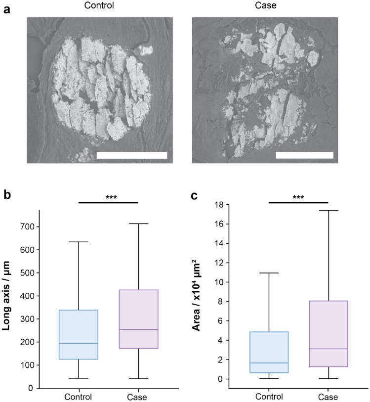
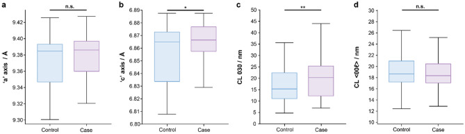
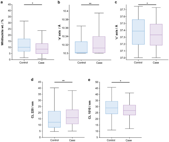
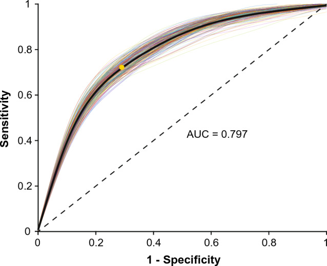
Ductal carcinoma in-situ (DCIS) accounts for 20-25% of all new breast cancer diagnoses. DCIS has an uncertain risk of progression to invasive breast cancer and a lack of predictive biomarkers may result in relatively high levels (~ 75%) of overtreatment. To identify unique prognostic biomarkers of invasive progression, crystallographic and chemical features of DCIS microcalcifications have been explored. Samples from patients with at least 5-years of follow up and no known recurrence (174 calcifications in 67 patients) or ipsilateral invasive breast cancer recurrence (179 microcalcifications in 57 patients) were studied. Significant differences were noted between the two groups including whitlockite relative mass, hydroxyapatite and whitlockite crystal maturity and, elementally, sodium to calcium ion ratio. A preliminary predictive model for DCIS to invasive cancer progression was developed from these parameters with an AUC of 0.797. These results provide insights into the differing DCIS tissue microenvironments, and how these impact microcalcification formation.

摘要

导管原位癌(DCIS)占所有新诊断乳腺癌的 20-25%。DCIS 向浸润性乳腺癌进展的风险不确定,缺乏预测性生物标志物可能导致过度治疗的比例相对较高(约 75%)。为了确定浸润性进展的独特预后生物标志物,已经探索了 DCIS 微钙化的晶体学和化学特征。对至少有 5 年随访且无已知复发(67 例患者中有 174 个钙化灶)或同侧浸润性乳腺癌复发(57 例患者中有 179 个微钙化灶)的患者进行了研究。两组之间存在显著差异,包括羟磷灰石相对质量、羟基磷灰石和羟磷灰石晶体成熟度以及元素钠钙离子比。从这些参数中开发了一个用于预测 DCIS 向浸润性癌进展的初步预测模型,AUC 为 0.797。这些结果提供了对不同 DCIS 组织微环境的深入了解,以及这些微环境如何影响微钙化的形成。

https://cdn.ncbi.nlm.nih.gov/pmc/blobs/2331/10250538/2715db202663/41598_2023_33547_Fig6_HTML.jpg
https://cdn.ncbi.nlm.nih.gov/pmc/blobs/2331/10250538/b62570900c49/41598_2023_33547_Fig1_HTML.jpg
https://cdn.ncbi.nlm.nih.gov/pmc/blobs/2331/10250538/3a5d5db7626d/41598_2023_33547_Fig2_HTML.jpg
https://cdn.ncbi.nlm.nih.gov/pmc/blobs/2331/10250538/80bbd98091a0/41598_2023_33547_Fig3_HTML.jpg
https://cdn.ncbi.nlm.nih.gov/pmc/blobs/2331/10250538/5a1d7849a692/41598_2023_33547_Fig4_HTML.jpg
https://cdn.ncbi.nlm.nih.gov/pmc/blobs/2331/10250538/48bfc9f98c02/41598_2023_33547_Fig5_HTML.jpg
https://cdn.ncbi.nlm.nih.gov/pmc/blobs/2331/10250538/2715db202663/41598_2023_33547_Fig6_HTML.jpg

相似文献

1
Microcalcification crystallography as a potential marker of DCIS recurrence.微钙化结晶学作为 DCIS 复发的潜在标志物。
Sci Rep. 2023 Jun 8;13(1):9331. doi: 10.1038/s41598-023-33547-8.
2
Qualitative Radiogenomics: Association Between BI-RADS Calcification Descriptors and Recurrence Risk as Assessed by the Oncotype DX Ductal Carcinoma In Situ Score.定性放射组学:BI-RADS 钙化描述符与 Oncotype DX 导管原位癌评分评估的复发风险之间的关联。
AJR Am J Roentgenol. 2019 Apr;212(4):919-924. doi: 10.2214/AJR.18.20306. Epub 2019 Feb 4.
3
Retrieval rate and accuracy of ultrasound-guided 14-G semi-automated core needle biopsy of breast microcalcifications.超声引导下 14-G 半自动核心针活检乳腺微钙化的检出率和准确性。
Korean J Radiol. 2014 Jan-Feb;15(1):12-9. doi: 10.3348/kjr.2014.15.1.12. Epub 2014 Jan 8.
4
Microcalcifications in 1657 Patients with Pure Ductal Carcinoma in Situ of the Breast: Correlation with Clinical, Histopathologic, Biologic Features, and Local Recurrence.1657例乳腺单纯导管原位癌患者的微钙化:与临床、组织病理学、生物学特征及局部复发的相关性
Ann Surg Oncol. 2016 Feb;23(2):482-9. doi: 10.1245/s10434-015-4876-6. Epub 2015 Sep 28.
5
Patterns of aggressiveness: risk of progression to invasive breast cancer by mammographic features of calcifications in screen-detected ductal carcinoma in situ.形态学侵袭性:乳腺 X 线筛查下原位导管癌的钙化形态特征与浸润性乳腺癌进展风险的关系。
Acta Radiol. 2022 May;63(5):586-595. doi: 10.1177/02841851211006319. Epub 2021 Apr 22.
6
Imaging Features of Patients Undergoing Active Surveillance for Ductal Carcinoma in Situ.行导管原位癌主动监测患者的影像学特征。
Acad Radiol. 2017 Nov;24(11):1364-1371. doi: 10.1016/j.acra.2017.05.017. Epub 2017 Jul 10.
7
Local tumor recurrence following breast-conservation therapy: correlation of histopathologic findings with detection method and mammographic findings.保乳治疗后局部肿瘤复发:组织病理学结果与检测方法及乳腺X线摄影结果的相关性
Radiology. 1999 Sep;212(3):829-35. doi: 10.1148/radiology.212.3.r99se41829.
8
A retrospective alternative for active surveillance trials for ductal carcinoma in situ of the breast.乳腺导管原位癌主动监测试验的回顾性替代方法。
Int J Cancer. 2020 Mar 1;146(5):1189-1197. doi: 10.1002/ijc.32362. Epub 2019 May 8.
9
Stromal architecture and periductal decorin are potential prognostic markers for ipsilateral locoregional recurrence in ductal carcinoma in situ of the breast.间质结构和管周decorin 可能是乳腺导管原位癌同侧局部区域复发的潜在预后标志物。
Histopathology. 2013 Oct;63(4):520-33. doi: 10.1111/his.12188. Epub 2013 Jul 26.
10
The role of stromal immune microenvironment in the progression of ductal carcinoma in situ (DCIS) to invasive breast cancer.基质免疫微环境在导管原位癌(DCIS)进展为浸润性乳腺癌中的作用。
Breast Cancer Res. 2021 Dec 24;23(1):118. doi: 10.1186/s13058-021-01494-9.

引用本文的文献

1
Vitacrystallography: Appearance and Development of Cancer-Induced Structural Biomarkers in a Mouse Model.维生素晶体学:小鼠模型中癌症诱导的结构生物标志物的出现与发展
Life (Basel). 2025 Jun 3;15(6):904. doi: 10.3390/life15060904.
2
Prostate microcalcification crystallography as a marker of pathology.前列腺微钙化晶体学作为病理学标志物
Sci Rep. 2025 Apr 29;15(1):14979. doi: 10.1038/s41598-025-98692-8.
3
Rapid Hyperspectral Photothermal Mid-Infrared Spectroscopic Imaging from Sparse Data for Gynecologic Cancer Tissue Subtyping.

本文引用的文献

1
A multi-modal exploration of heterogeneous physico-chemical properties of DCIS breast microcalcifications.多模态探索 DCIS 乳腺微钙化的异质物理化学特性。
Analyst. 2022 Apr 11;147(8):1641-1654. doi: 10.1039/d1an01548f.
2
Breast microcalcifications: Past, present and future (Review).乳腺微钙化:过去、现在与未来(综述)
Mol Clin Oncol. 2022 Apr;16(4):81. doi: 10.3892/mco.2022.2514. Epub 2022 Feb 10.
3
Low-risk DCIS. What is it? Observe or excise?低危 DCIS。这是什么?观察还是切除?
基于稀疏数据的妇科癌症组织亚型快速高光谱光热中红外光谱成像。
Anal Chem. 2024 Oct 8;96(40):15880-15887. doi: 10.1021/acs.analchem.4c01093. Epub 2024 Sep 23.
4
Characterization of dolomite and calcite microcalcifications in human breast tissue.人体乳腺组织中白云石和方解石微钙化的表征
RSC Adv. 2024 Sep 10;14(39):28741-28752. doi: 10.1039/d4ra04137b. eCollection 2024 Sep 4.
5
Assessing the malignancy of suspicious breast microcalcifications: the role of contrast enhanced mammography.评估可疑乳腺微钙化的恶性程度:对比增强乳腺摄影的作用。
Radiol Med. 2024 Jun;129(6):855-863. doi: 10.1007/s11547-024-01813-y. Epub 2024 Apr 12.
6
Translating microcalcification biomarker information into the laboratory: A preliminary assessment utilizing core biopsies obtained from sites of mammographic calcification.将微钙化生物标志物信息转化到实验室:利用从乳腺钼靶钙化部位获取的芯针活检进行的初步评估。
Heliyon. 2024 Mar 11;10(6):e27686. doi: 10.1016/j.heliyon.2024.e27686. eCollection 2024 Mar 30.
7
Rapid hyperspectral photothermal mid-infrared spectroscopic imaging from sparse data for gynecologic cancer tissue subtyping.基于稀疏数据的快速高光谱光热中红外光谱成像用于妇科癌组织亚型分析
ArXiv. 2024 Feb 28:arXiv:2402.17960v1.
Virchows Arch. 2022 Jan;480(1):21-32. doi: 10.1007/s00428-021-03173-8. Epub 2021 Aug 27.
4
Long-term risk of subsequent ipsilateral lesions after surgery with or without radiotherapy for ductal carcinoma in situ of the breast.乳腺导管原位癌保乳手术或不保乳手术后同侧病变的长期风险。
Br J Cancer. 2021 Nov;125(10):1443-1449. doi: 10.1038/s41416-021-01496-6. Epub 2021 Aug 18.
5
Mammographic microcalcifications and risk of breast cancer.乳腺钼靶微钙化与乳腺癌风险。
Br J Cancer. 2021 Aug;125(5):759-765. doi: 10.1038/s41416-021-01459-x. Epub 2021 Jun 14.
6
A time-course Raman spectroscopic analysis of spontaneous in vitro microcalcifications in a breast cancer cell line.对乳腺癌细胞系中自发的体外微钙化进行时程拉曼光谱分析。
Lab Invest. 2021 Sep;101(9):1267-1280. doi: 10.1038/s41374-021-00619-0. Epub 2021 Jun 11.
7
Calcium signaling: breast cancer's approach to manipulation of cellular circuitry.钙信号传导:乳腺癌对细胞信号通路的操控方式
Biophys Rev. 2020 Dec 18;12(6):1343-1359. doi: 10.1007/s12551-020-00771-9. eCollection 2020 Dec.
8
The harsh microenvironment in early breast cancer selects for a Warburg phenotype.早期乳腺癌恶劣的微环境选择出瓦博格表型。
Proc Natl Acad Sci U S A. 2021 Jan 19;118(3). doi: 10.1073/pnas.2011342118.
9
Multiple Pathways for Pathological Calcification in the Human Body.人体病理性钙化的多种途径。
Adv Healthc Mater. 2021 Feb;10(4):e2001271. doi: 10.1002/adhm.202001271. Epub 2020 Dec 4.
10
Mixed-Methods Study to Predict Upstaging of DCIS to Invasive Disease on Mammography.混合方法研究预测 DCIS 在乳腺 X 线摄影中向浸润性疾病的升级。
AJR Am J Roentgenol. 2021 Apr;216(4):903-911. doi: 10.2214/AJR.20.23679. Epub 2021 Feb 3.