Li Chaohong, Liu Fengting, Li Xingyu, Yang Xiaohua, Li Qiaoqiao, Huang Jing
Department of Cardiology, The Second Affiliated Hospital of Chongqing Medical University, Chongqing, China.
Henan Key Laboratory of Neurorestoratology, Life Science Research Center, The First Affiliated Hospital of Xinxiang Medical University, Weihui, Henan, China.
BMC Genomics. 2025 Apr 29;26(1):421. doi: 10.1186/s12864-025-11559-0.
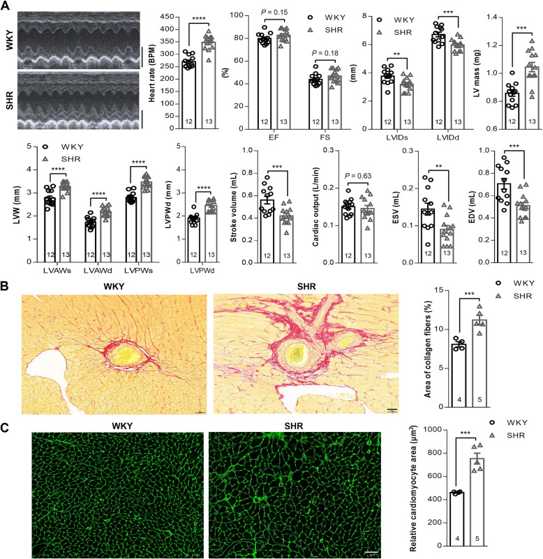
Hypertension-induced left ventricular hypertrophy (LVH) is a cardiac structural remodeling and dysfunction resulting from chronic hypertension and is an independent risk factor for cardiovascular morbidity and mortality. Studies have implicated the involvement of the adrenal gland (AG) and superior cervical ganglion (SCG) in hypertension regulation. However, the molecular mechanisms of AG and SCG during hypertension-induced LVH remain unclear. In this study, we investigated the transcriptome characteristics of the left ventricle, AG, and SCG in 24-week-old spontaneously hypertensive rats (SHR) and Wistar-Kyoto (WKY) rats using transcriptome sequencing (RNA-seq) technology.
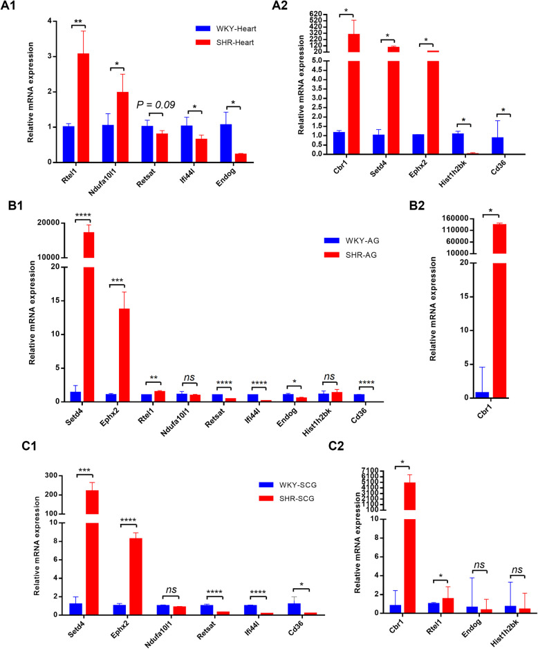
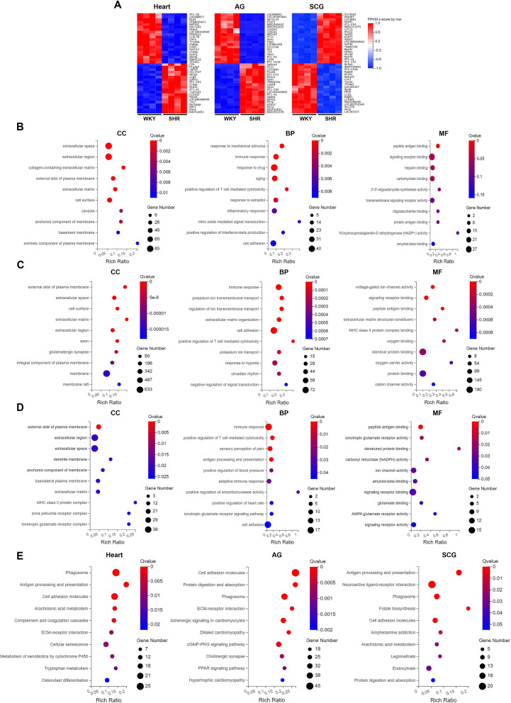
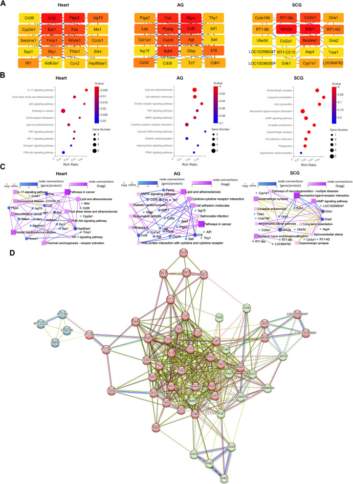
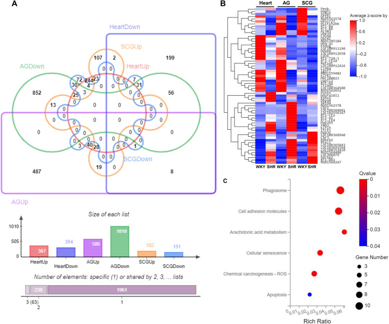
We identified 671 differentially expressed genes (DEGs) in the left ventricle, 1596 DEGs in the AG, and 343 DEGs in the SCG between SHR and WKY rats. Functional analysis revealed that these DEGs were involved in pathways related to immune inflammation, cellular senescence, metabolic responses, and synaptic transmission. We also identified the top 20 hub genes specific to each of the three organs. PPI network analysis showed that these hub genes were highly interconnected and clustered into three functional modules. KEGG pathway analysis revealed that the hub genes in the left ventricle and AG were mainly enriched in inflammation-related pathways, while the hub genes in the SCG were primarily enriched in synapse-related pathways. Venn diagram analysis identified 63 overlapping DEGs across the three organs, with 59 DEGs exhibiting consistent expression changes.
In summary, our study elucidates the transcriptome features of AG and SCG during hypertension-induced LVH in SHR.
高血压诱导的左心室肥厚(LVH)是一种由慢性高血压导致的心脏结构重塑和功能障碍,是心血管疾病发病和死亡的独立危险因素。研究表明肾上腺(AG)和颈上神经节(SCG)参与高血压调节。然而,在高血压诱导的LVH过程中AG和SCG的分子机制仍不清楚。在本研究中,我们使用转录组测序(RNA-seq)技术研究了24周龄自发性高血压大鼠(SHR)和Wistar-Kyoto(WKY)大鼠左心室、AG和SCG的转录组特征。
我们在SHR和WKY大鼠之间的左心室中鉴定出671个差异表达基因(DEG),在AG中鉴定出1596个DEG,在SCG中鉴定出343个DEG。功能分析表明,这些DEG参与了与免疫炎症、细胞衰老、代谢反应和突触传递相关的途径。我们还确定了三个器官各自特有的前20个枢纽基因。蛋白质-蛋白质相互作用(PPI)网络分析表明,这些枢纽基因高度互联并聚集成三个功能模块。京都基因与基因组百科全书(KEGG)通路分析表明,左心室和AG中的枢纽基因主要富集于炎症相关通路,而SCG中的枢纽基因主要富集于突触相关通路。维恩图分析确定了三个器官中63个重叠的DEG,其中59个DEG表现出一致的表达变化。
总之,我们的研究阐明了SHR中高血压诱导的LVH过程中AG和SCG的转录组特征。