• 文献检索
  • 文档翻译
  • 深度研究
  • 学术资讯
  • Suppr Zotero 插件Zotero 插件
  • 邀请有礼
  • 套餐&价格
  • 历史记录
应用&插件
Suppr Zotero 插件Zotero 插件浏览器插件Mac 客户端Windows 客户端微信小程序
定价
高级版会员购买积分包购买API积分包
服务
文献检索文档翻译深度研究API 文档MCP 服务
关于我们
关于 Suppr公司介绍联系我们用户协议隐私条款
关注我们

Suppr 超能文献

核心技术专利:CN118964589B侵权必究
粤ICP备2023148730 号-1Suppr @ 2026

文献检索

告别复杂PubMed语法,用中文像聊天一样搜索,搜遍4000万医学文献。AI智能推荐,让科研检索更轻松。

立即免费搜索

文件翻译

保留排版,准确专业,支持PDF/Word/PPT等文件格式,支持 12+语言互译。

免费翻译文档

深度研究

AI帮你快速写综述,25分钟生成高质量综述,智能提取关键信息,辅助科研写作。

立即免费体验

肝脏良性局灶性病变超声造影剂的洗脱——频率及可能原因综述

Washout on Contrast-Enhanced Ultrasound of Benign Focal Liver Lesions-A Review on Its Frequency and Possible Causes.

作者信息

Möller Kathleen, Görg Christian, Krix Martin, Jenssen Christian, Dong Yi, Cui Xin-Wu, Dietrich Christoph F

机构信息

Medical Department I/Gastroenterology, SANA Hospital Lichtenberg, 10365 Berlin, Germany.

Interdisciplinary Center of Ultrasound Diagnostics, Gastroenterology, Endocrinology, Metabolism and Clinical Infectiology, University Hospital Giessen and Marburg, Philipp University of Marburg, Baldingerstraße, 35037 Marburg, Germany.

出版信息

Diagnostics (Basel). 2025 Apr 14;15(8):998. doi: 10.3390/diagnostics15080998.

DOI:10.3390/diagnostics15080998
PMID:40310346
原文链接:https://pmc.ncbi.nlm.nih.gov/articles/PMC12025567/
Abstract

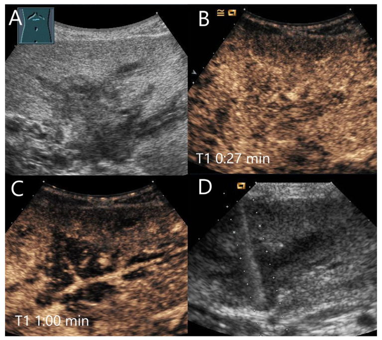
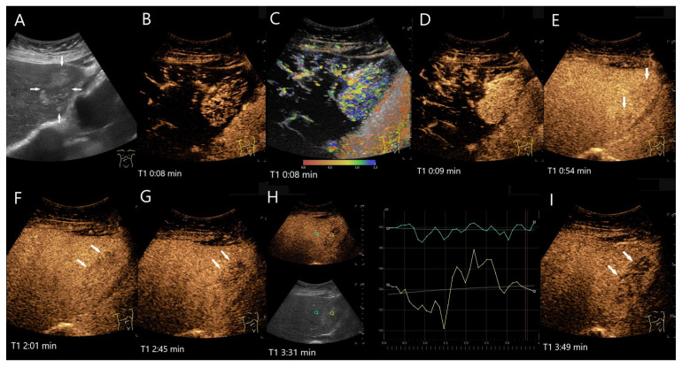
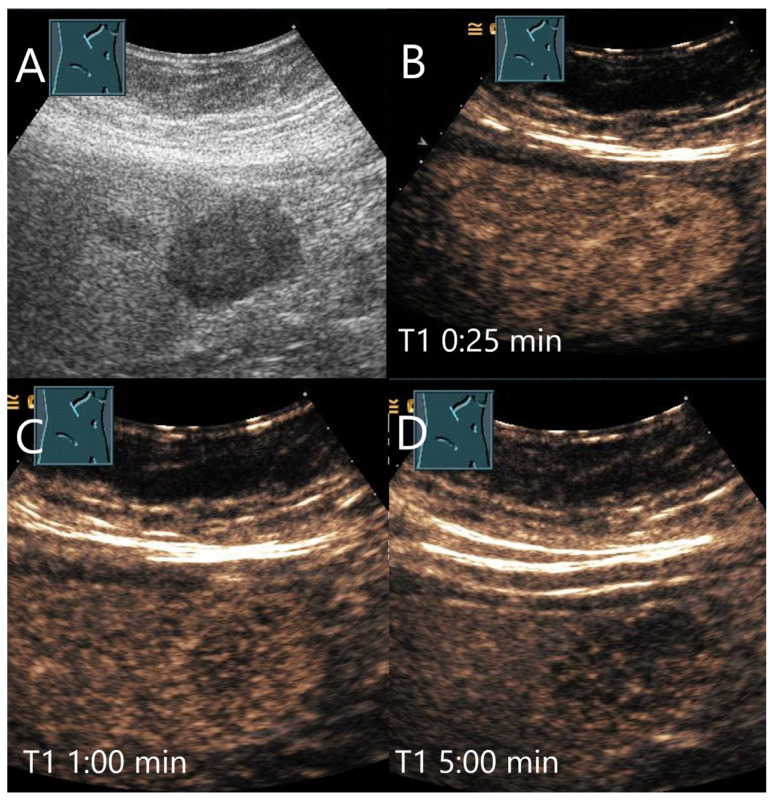
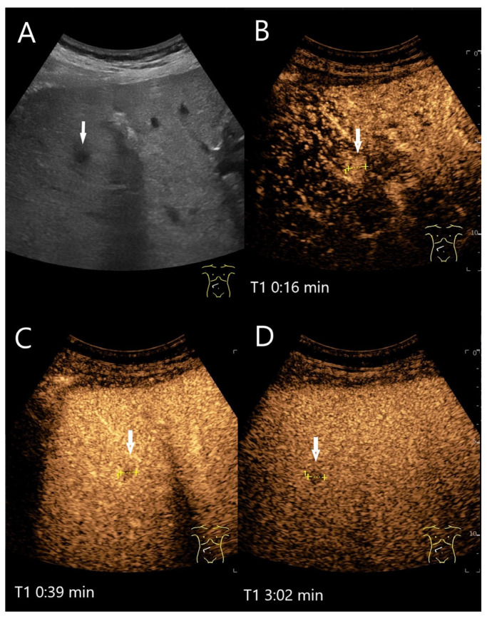
In all imaging methods, including contrast-enhanced ultrasound (CEUS), enhancement in the late phase (LP) is an important criterion for differentiating between benign and malignant focal liver lesions (FLLs). In general, malignant liver lesions are characterized by hypoenhancement and washout in the LP. A lesion with LP hyperenhancement or isoenhancement in the non-cirrhotic liver is usually benign. However, LP hypoenhancement in benign lesions is not so rare, and is even normal and the standard for some lesions, and there are exceptions for each tumor entity that can represent a diagnostic challenge. Knowing these contrast patterns and exceptions is key for correct diagnosis and patient management. The following narrative review describes the contrast behaviors and the frequency of washout and LP hypoenhancement for common as well as rare benign liver lesions and analyzes its causes.

摘要

在包括超声造影(CEUS)在内的所有成像方法中,延迟期(LP)强化是鉴别肝脏局灶性病变(FLLs)良恶性的重要标准。一般来说,恶性肝脏病变的特征是延迟期低强化和廓清。在非肝硬化肝脏中,延迟期高强化或等强化的病变通常为良性。然而,良性病变中延迟期低强化并不罕见,甚至对某些病变来说是正常表现和标准,而且每个肿瘤实体都有例外情况,这可能带来诊断挑战。了解这些造影模式和例外情况是正确诊断和患者管理的关键。以下叙述性综述描述了常见及罕见良性肝脏病变的造影表现、廓清频率和延迟期低强化情况,并分析其原因。

https://cdn.ncbi.nlm.nih.gov/pmc/blobs/4b41/12025567/543b259471f3/diagnostics-15-00998-g023.jpg
https://cdn.ncbi.nlm.nih.gov/pmc/blobs/4b41/12025567/46cafcfaf6fc/diagnostics-15-00998-g001.jpg
https://cdn.ncbi.nlm.nih.gov/pmc/blobs/4b41/12025567/90b45645a236/diagnostics-15-00998-g002.jpg
https://cdn.ncbi.nlm.nih.gov/pmc/blobs/4b41/12025567/f73a26757c45/diagnostics-15-00998-g003.jpg
https://cdn.ncbi.nlm.nih.gov/pmc/blobs/4b41/12025567/11cc1943ec59/diagnostics-15-00998-g004.jpg
https://cdn.ncbi.nlm.nih.gov/pmc/blobs/4b41/12025567/108c6c20d25a/diagnostics-15-00998-g005.jpg
https://cdn.ncbi.nlm.nih.gov/pmc/blobs/4b41/12025567/3fafcb720a30/diagnostics-15-00998-g006.jpg
https://cdn.ncbi.nlm.nih.gov/pmc/blobs/4b41/12025567/c35594624618/diagnostics-15-00998-g007.jpg
https://cdn.ncbi.nlm.nih.gov/pmc/blobs/4b41/12025567/a1475166e120/diagnostics-15-00998-g008.jpg
https://cdn.ncbi.nlm.nih.gov/pmc/blobs/4b41/12025567/4b89d3754b58/diagnostics-15-00998-g009.jpg
https://cdn.ncbi.nlm.nih.gov/pmc/blobs/4b41/12025567/3be6b3885872/diagnostics-15-00998-g010.jpg
https://cdn.ncbi.nlm.nih.gov/pmc/blobs/4b41/12025567/55c77d4524dd/diagnostics-15-00998-g011.jpg
https://cdn.ncbi.nlm.nih.gov/pmc/blobs/4b41/12025567/204fa159bc39/diagnostics-15-00998-g012.jpg
https://cdn.ncbi.nlm.nih.gov/pmc/blobs/4b41/12025567/3b86cccb3539/diagnostics-15-00998-g013.jpg
https://cdn.ncbi.nlm.nih.gov/pmc/blobs/4b41/12025567/a1b001a716a9/diagnostics-15-00998-g014.jpg
https://cdn.ncbi.nlm.nih.gov/pmc/blobs/4b41/12025567/8e6888f2e90d/diagnostics-15-00998-g015.jpg
https://cdn.ncbi.nlm.nih.gov/pmc/blobs/4b41/12025567/1a9e1ab2d496/diagnostics-15-00998-g016.jpg
https://cdn.ncbi.nlm.nih.gov/pmc/blobs/4b41/12025567/e188740a484e/diagnostics-15-00998-g017.jpg
https://cdn.ncbi.nlm.nih.gov/pmc/blobs/4b41/12025567/8728368ddf9e/diagnostics-15-00998-g018.jpg
https://cdn.ncbi.nlm.nih.gov/pmc/blobs/4b41/12025567/9fcbd96bb25e/diagnostics-15-00998-g019.jpg
https://cdn.ncbi.nlm.nih.gov/pmc/blobs/4b41/12025567/c8eaadacfe16/diagnostics-15-00998-g020.jpg
https://cdn.ncbi.nlm.nih.gov/pmc/blobs/4b41/12025567/2dddef26cc65/diagnostics-15-00998-g021.jpg
https://cdn.ncbi.nlm.nih.gov/pmc/blobs/4b41/12025567/4ecc865461b5/diagnostics-15-00998-g022.jpg
https://cdn.ncbi.nlm.nih.gov/pmc/blobs/4b41/12025567/543b259471f3/diagnostics-15-00998-g023.jpg

相似文献

1
Washout on Contrast-Enhanced Ultrasound of Benign Focal Liver Lesions-A Review on Its Frequency and Possible Causes.肝脏良性局灶性病变超声造影剂的洗脱——频率及可能原因综述
Diagnostics (Basel). 2025 Apr 14;15(8):998. doi: 10.3390/diagnostics15080998.
2
Contrast-enhanced ultrasound of hepatocarcinogenesis in liver cirrhosis.肝硬化肝癌发生的超声造影研究。
Chin Med J (Engl). 2012 Sep;125(17):3104-9.
3
Imaging Features of Hepatocellular Carcinoma in the Non-Cirrhotic Liver with Sonazoid-Enhanced Contrast-Enhanced Ultrasound.使用Sonazoid增强型超声造影对非肝硬化肝脏中肝细胞癌的成像特征
Diagnostics (Basel). 2022 Sep 20;12(10):2272. doi: 10.3390/diagnostics12102272.
4
Tumor-specific vascularization pattern of liver metastasis, hepatocellular carcinoma, hemangioma and focal nodular hyperplasia in the differential diagnosis of 1,349 liver lesions in contrast-enhanced ultrasound (CEUS).对比增强超声(CEUS)对1349例肝脏病变进行鉴别诊断时,肝转移瘤、肝细胞癌、肝血管瘤和局灶性结节性增生的肿瘤特异性血管生成模式
Ultraschall Med. 2009 Aug;30(4):376-82. doi: 10.1055/s-0028-1109672. Epub 2009 Aug 17.
5
Strategy for Accurate Diagnosis by Contrast-Enhanced Ultrasound of Focal Liver Lesions in Patients Not at High Risk for Hepatocellular Carcinoma: A Preliminary Study.对比增强超声在非高危人群肝脏局灶性病变中的准确诊断策略:一项初步研究。
J Ultrasound Med. 2023 Jun;42(6):1333-1344. doi: 10.1002/jum.16151. Epub 2022 Dec 19.
6
Characterization of Focal Liver Lesions Indistinctive on B Mode Ultrasound: Benefits of Contrast-Enhanced Ultrasound.B 型超声检查无特征性的局灶性肝病变的特征:超声造影的优势
Biomed Res Int. 2017;2017:8970156. doi: 10.1155/2017/8970156. Epub 2017 Apr 11.
7
Contrast-Enhanced Ultrasound With Perfluorobutane for Hepatocellular Carcinoma Diagnosis: Comparison of Imaging Phases and Diagnostic Criteria.对比增强超声联合全氟丁烷在肝细胞癌诊断中的应用:成像相和诊断标准的比较。
AJR Am J Roentgenol. 2024 Feb;222(2):e2330156. doi: 10.2214/AJR.23.30156. Epub 2023 Nov 22.
8
Contrast-enhanced ultrasound using SonoVue® (sulphur hexafluoride microbubbles) compared with contrast-enhanced computed tomography and contrast-enhanced magnetic resonance imaging for the characterisation of focal liver lesions and detection of liver metastases: a systematic review and cost-effectiveness analysis.超声造影使用声诺维®(六氟化硫微泡)与对比增强计算机断层扫描和对比增强磁共振成像在局灶性肝脏病变的特征描述和肝转移检测中的比较:系统评价和成本效益分析。
Health Technol Assess. 2013 Apr;17(16):1-243. doi: 10.3310/hta17160.
9
Development and validation of an ultrasound-based prediction model for differentiating between malignant and benign solid pancreatic lesions.基于超声的区分胰腺良恶性实性病灶的预测模型的建立与验证。
Eur Radiol. 2022 Dec;32(12):8296-8305. doi: 10.1007/s00330-022-08930-0. Epub 2022 Jun 25.
10
Contrast-enhanced ultrasound with microbubbles containing sulfur hexafluoride and perfluorobutane with Kupffer phase for the detection of colorectal liver metastases.含六氟化硫和全氟丁烷的微泡超声造影与库普弗期在结直肠癌肝转移检测中的应用。
Eur Radiol. 2024 Jan;34(1):622-631. doi: 10.1007/s00330-023-10051-1. Epub 2023 Aug 11.

本文引用的文献

1
Clinical and Multimodal Imaging Features of Hepatic Inflammatory Pseudotumors: A Two-Center Retrospective Study.肝脏炎性假瘤的临床及多模态影像学特征:一项双中心回顾性研究
J Ultrasound Med. 2025 Apr;44(4):691-701. doi: 10.1002/jum.16629. Epub 2024 Dec 12.
2
Ceus features of liver pecoma: a case report and literature review.肝脏血管周上皮样细胞瘤的超声特征:1例报告并文献复习
J Ultrasound. 2025 Mar;28(1):261-268. doi: 10.1007/s40477-024-00973-x. Epub 2024 Nov 18.
3
Comparative efficacy of contrast-enhanced ultrasound versus B-mode ultrasound in the diagnosis and monitoring of hepatic abscesses.
超声造影与B超在肝脓肿诊断及监测中的疗效比较
Pol J Radiol. 2024 Oct 8;89:e470-e479. doi: 10.5114/pjr/192184. eCollection 2024.
4
Contrast-enhanced ultrasound features of hepatic angiomyolipoma: comparison with AFP-negative and non-viral hepatocellular carcinoma.肝脏血管平滑肌脂肪瘤的超声造影特征:与甲胎蛋白阴性及非病毒性肝细胞癌的比较
Ultrasound Int Open. 2024 Jun 14;10:a23186654. doi: 10.1055/a-2318-6654. eCollection 2024.
5
Utility of Contrast-Enhanced Ultrasound in Optimizing Hepatic Abscess Treatment and Monitoring.超声造影在优化肝脓肿治疗及监测中的应用价值
J Clin Med. 2024 Aug 26;13(17):5046. doi: 10.3390/jcm13175046.
6
Hepatic perivascular epithelioid cell tumor: a retrospective analysis of 36 cases.肝血管周上皮样细胞肿瘤:36例回顾性分析
Front Oncol. 2024 Aug 16;14:1416254. doi: 10.3389/fonc.2024.1416254. eCollection 2024.
7
Comments and illustrations of the WFUMB CEUS liver guidelines: rare benign hematological focal liver lesions (hepatic extramedullary hematopoiesis, Hemophagocytic lymphohistiocytosis, reactive lymphoid hyperplasia).WFUMB 超声造影肝脏指南的评论与说明:罕见的良性血液系统局灶性肝脏病变(肝外髓外造血、噬血细胞性淋巴组织细胞增生症、反应性淋巴样增生)
Med Ultrason. 2025 Mar 2;27(1):73-81. doi: 10.11152/mu-4419. Epub 2024 Aug 10.
8
The value of histopathologic examination and Xpert (MTB/RIF) assay in diagnosis of cervical lymph node tuberculosis after coarse needle biopsy guided by CEUS: a retrospective analysis of 612 cases.超声造影引导下粗针穿刺活检后组织病理学检查及Xpert(MTB/RIF)检测在颈部淋巴结结核诊断中的价值:612例回顾性分析
Eur J Clin Microbiol Infect Dis. 2024 Oct;43(10):1951-1957. doi: 10.1007/s10096-024-04913-9. Epub 2024 Aug 1.
9
Granulomatous liver diseases.肉芽肿性肝病。
Hepatol Commun. 2024 Mar 18;8(4). doi: 10.1097/HC9.0000000000000392. eCollection 2024 Apr 1.
10
The Effect of Contrast-enhanced Ultrasound Vessels and Surgical Drains Guidance Percutaneous Catheter Drainage in the Treatment of Pyogenic Liver Abscess.超声造影血管及手术引流管引导经皮置管引流在化脓性肝脓肿治疗中的作用
Curr Med Imaging. 2024 Mar 7. doi: 10.2174/0115734056261616231224161652.