Department of Ultrasound, West China Hospital of Sichuan University, Chengdu, 610041, China.
Department of Ultrasound, Tianjin Third Central Hospital, Tianjin, 300170, China.
Eur Radiol. 2022 Dec;32(12):8296-8305. doi: 10.1007/s00330-022-08930-0. Epub 2022 Jun 25.
To identify the diagnostic ability of precontrast and contrast-enhanced ultrasound (CEUS) in differentiating between malignant and benign solid pancreatic lesions (MSPLs and BSPLs) and to develop an easy-to-use diagnostic nomogram.
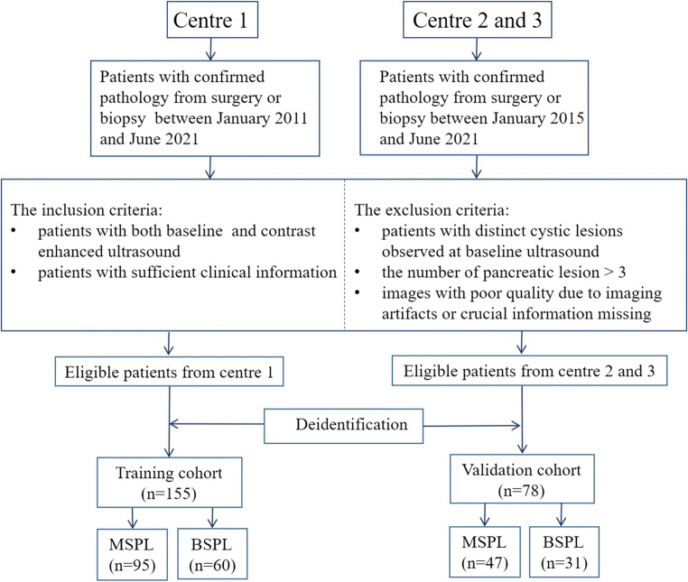
This study was approved by the institutional review board. Patients with pathologically confirmed solid pancreatic lesions were enrolled from one tertiary medical centre from March 2011 to June 2021 and in two tertiary institutions between January 2015 and June 2021. A prediction nomogram model was established in the training set by using precontrast US and CEUS imaging features that were independently associated with MSPLs. The performance of the prediction model was further externally validated.
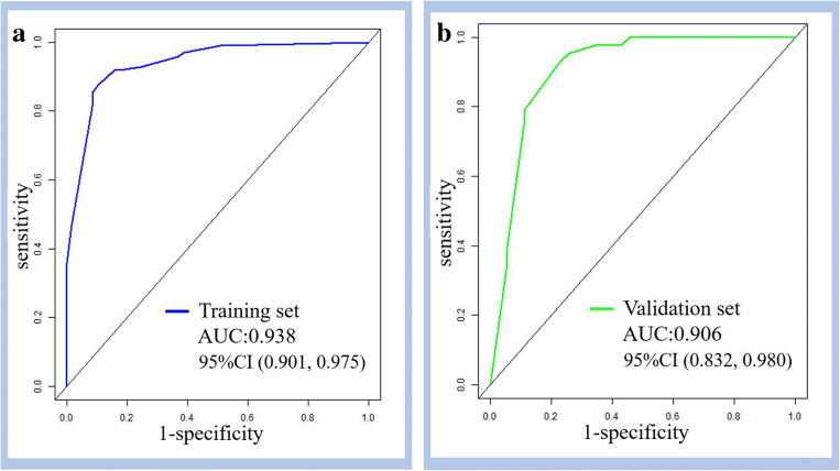
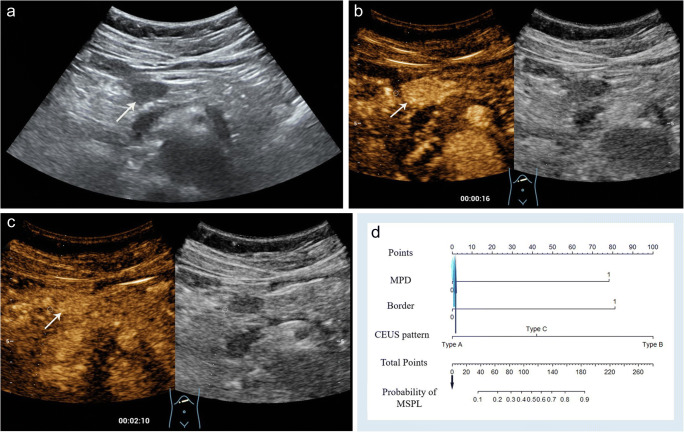
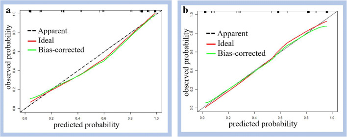
A total of 155 patients (mean age, 55 ± 14.6 years, M/F = 84/71) and 78 patients (mean age, 59 ± 13.4 years, M/F = 36/42) were included in the training and validation cohorts, respectively. In the training set, an ill-defined border and dilated main pancreatic duct on precontrast ultrasound, CEUS patterns of hypoenhancement in both the arterial and venous phases of CEUS, and hyperenhancement/isoenhancement followed by washout were independently associated with MSPLs. The prediction nomogram model developed with the aforementioned variables showed good performance in differentiating MSPLs from BSPLs with an area under the curve (AUC) of 0.938 in the training set and 0.906 in the validation set.
Hypoenhancement in all phases, hyperenhancement/isoenhancement followed by washout on CEUS, an ill-defined border, and a dilated main pancreatic duct were independent risk factors for MSPLs. The nomogram constructed based on these predictors can be used to diagnose MSPLs.
• An ill-defined border and dilated main pancreatic duct on precontrast ultrasound, hypoenhancement in all phases of CEUS, and hyperenhancement/isoenhancement followed by washout were independently associated with MSPLs. • The ultrasound-based prediction model showed good performance in differentiating MSPLs from BSPLs with an AUC of 0.938 in the training set and 0.906 in the external validation set. • An ultrasound-based nomogram is an easy-to-use tool to differentiate between MSPLs and BSPLs with high efficacy.
确定超声造影前和造影增强超声(CEUS)在鉴别良恶性胰腺实性病变(MSPL 和 BSPL)中的诊断能力,并建立一个易于使用的诊断列线图。
本研究经机构审查委员会批准。2011 年 3 月至 2021 年 6 月,从一家三级医疗中心招募了经病理证实的胰腺实性病变患者,2015 年 1 月至 2021 年 6 月,从两家三级医疗机构招募了经病理证实的胰腺实性病变患者。通过使用与 MSPL 独立相关的超声造影前和 CEUS 成像特征,在训练集中建立预测列线图模型。进一步对预测模型的性能进行外部验证。
在训练组中,共纳入 155 例患者(平均年龄 55±14.6 岁,M/F=84/71)和 78 例患者(平均年龄 59±13.4 岁,M/F=36/42)。在训练集中,超声造影前边界不清晰和主胰管扩张、CEUS 动脉期和静脉期均呈低增强、增强/等增强后洗脱为 MSPL 的独立危险因素。使用上述变量建立的预测列线图模型在训练集和验证集区分 MSPL 和 BSPL 的曲线下面积(AUC)分别为 0.938 和 0.906,表现出良好的性能。
CEUS 各期低增强、增强/等增强后洗脱、边界不清晰和主胰管扩张为 MSPL 的独立危险因素。基于这些预测因素建立的列线图可用于诊断 MSPL。
超声造影前边界不清晰和主胰管扩张、CEUS 各期低增强、增强/等增强后洗脱与 MSPL 独立相关。
基于超声的预测模型在训练集和外部验证集中区分 MSPL 和 BSPL 的 AUC 分别为 0.938 和 0.906,具有良好的性能。
基于超声的列线图是一种易于使用的工具,可有效区分 MSPL 和 BSPL。