Sultan Nessma, Mowafey Bassant, Ata Fatma, El-Zekrid Mona H, Jayash Soher Nagi
Faculty of Dentistry, Mansoura University, Mansoura, Egypt; Faculty of Dentistry, Mansoura National University, Gamasa, Egypt.
Faculty of Dentistry, Mansoura University, Egypt.
Int Dent J. 2025 May 3;75(4):100817. doi: 10.1016/j.identj.2025.03.026.
Alveolar bone resorption following tooth extraction presents significant challenges for implant-supported rehabilitations. Demineralised dentin matrix (DDM) has emerged as a promising scaffold for bone tissue regeneration. This study evaluates the bone-regenerating potential of varying degrees of dentin demineralisation.
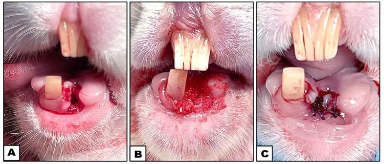
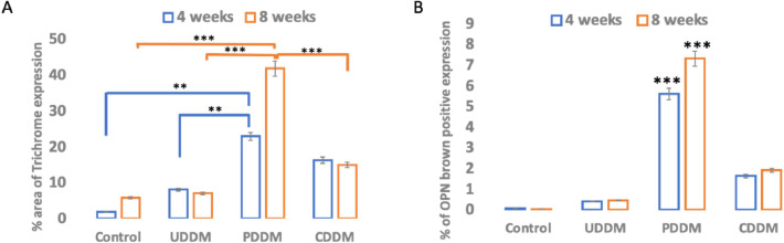
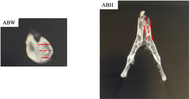
Thirty-two male white New Zealand rabbits underwent extraction of the left mandibular anterior tooth and were assigned to 4 groups: undemineralised dentin matrix (UDDM), partially demineralised dentin matrix (PDDM), completely demineralised dentin matrix (CDDM), and a control group with no treatment. At 4 and 8 weeks post extraction, cone-beam computed tomography (CBCT) was used to assess alveolar bone height and width. Histological analyses using H&E and Masson trichrome stains evaluated new bone formation, and immunohistochemistry detected osteopontin expression.
CBCT imaging revealed progressive increases in alveolar bone height and width across all groups over time. Histological analysis showed new bone formation in all groups, with the PDDM group demonstrating closer integration of newly formed bone trabeculae compared with the others. IHC results showed higher osteopontin expression in the PDDM group, highlighting its superior bone-inductive potential.
Among the tested materials, PDDM exhibited the most effective bone induction and tissue regeneration capabilities, outperforming CDDM and UDDM in promoting alveolar bone repair. These findings position PDDM as a valuable scaffold for enhancing bone tissue regeneration in clinical applications.
The use of PDDM in tooth extraction sockets significantly promotes efficient and reliable bone regeneration, making it a valuable option for clinical applications in implant dentistry.
拔牙后牙槽骨吸收给种植修复带来了重大挑战。脱矿牙本质基质(DDM)已成为骨组织再生的一种有前景的支架材料。本研究评估不同程度牙本质脱矿的骨再生潜力。
32只雄性白色新西兰兔拔除左下颌前牙,并分为4组:未脱矿牙本质基质(UDDM)组、部分脱矿牙本质基质(PDDM)组、完全脱矿牙本质基质(CDDM)组和未治疗的对照组。拔牙后4周和8周,使用锥形束计算机断层扫描(CBCT)评估牙槽骨高度和宽度。采用苏木精-伊红(H&E)染色和马松三色染色进行组织学分析,评估新骨形成情况,免疫组织化学检测骨桥蛋白表达。
CBCT成像显示,随着时间推移,所有组的牙槽骨高度和宽度均逐渐增加。组织学分析显示所有组均有新骨形成,与其他组相比,PDDM组新形成的骨小梁整合更紧密。免疫组化结果显示PDDM组骨桥蛋白表达更高,突出了其优越的骨诱导潜力。
在测试材料中,PDDM表现出最有效的骨诱导和组织再生能力,在促进牙槽骨修复方面优于CDDM和UDDM。这些发现表明PDDM是临床应用中增强骨组织再生的一种有价值的支架材料。
在拔牙窝中使用PDDM可显著促进高效可靠的骨再生,使其成为种植牙科临床应用的一种有价值的选择。