• 文献检索
  • 文档翻译
  • 深度研究
  • 学术资讯
  • Suppr Zotero 插件Zotero 插件
  • 邀请有礼
  • 套餐&价格
  • 历史记录
应用&插件
Suppr Zotero 插件Zotero 插件浏览器插件Mac 客户端Windows 客户端微信小程序
定价
高级版会员购买积分包购买API积分包
服务
文献检索文档翻译深度研究API 文档MCP 服务
关于我们
关于 Suppr公司介绍联系我们用户协议隐私条款
关注我们

Suppr 超能文献

核心技术专利:CN118964589B侵权必究
粤ICP备2023148730 号-1Suppr @ 2026

文献检索

告别复杂PubMed语法,用中文像聊天一样搜索,搜遍4000万医学文献。AI智能推荐,让科研检索更轻松。

立即免费搜索

文件翻译

保留排版,准确专业,支持PDF/Word/PPT等文件格式,支持 12+语言互译。

免费翻译文档

深度研究

AI帮你快速写综述,25分钟生成高质量综述,智能提取关键信息,辅助科研写作。

立即免费体验

心肌球衍生细胞对大鼠苏金/低氧诱导的伴有右心室功能障碍的肺动脉高压模型中适应不良的右心室心肌的影响。

Impact of cardiosphere-derived cells on the maladapted right ventricular muscle in a rat sugen/hypoxia model of pulmonary hypertension with right ventricular dysfunction.

作者信息

Middleton Ryan C, Karpov Oleg A, Fournier Mario, Kreimer Simion, Mastali Mitra, Liu Weixin, Li Liang, Voelkel Norbert F, Van Eyk Jennifer E, Marbán Eduardo, Lewis Michael I

机构信息

Smidt Heart Institute, Cedars-Sinai Medical Center, Los Angeles, California, United States of America.

Amsterdam University Medical Centers, Amsterdam, Netherlands.

出版信息

PLoS One. 2025 May 12;20(5):e0321895. doi: 10.1371/journal.pone.0321895. eCollection 2025.

DOI:10.1371/journal.pone.0321895
PMID:40354360
原文链接:https://pmc.ncbi.nlm.nih.gov/articles/PMC12068596/
Abstract

BACKGROUND AND AIMS

With pulmonary arterial hypertension (PAH), right ventricular (RV) function is a major determinant of survival. Despite current therapies, maladaptive changes ensue in the RV muscle of PAH patients, culminating in RV dysfunction and failure. The aims of the study were to evaluate the impact of intra-coronary (IC) cardiosphere-derived cells (CDCs) in attenuating the maladaptive pathobiology in the RV muscle and evaluating mechanisms underlying improvements in RV function.

METHODS

Two groups of the Sugen/Hypoxia rat model of PAH, exhibiting significantly reduced RV function, via TAPSE measurements, received either intracoronary infusion of CDCs or PBS placebo. Immunohistochemistry methods were used to assess RV pathobiological changes. Additionally, advanced proteomics were employed to examine protein signaling pathways and upstream regulators.

RESULTS

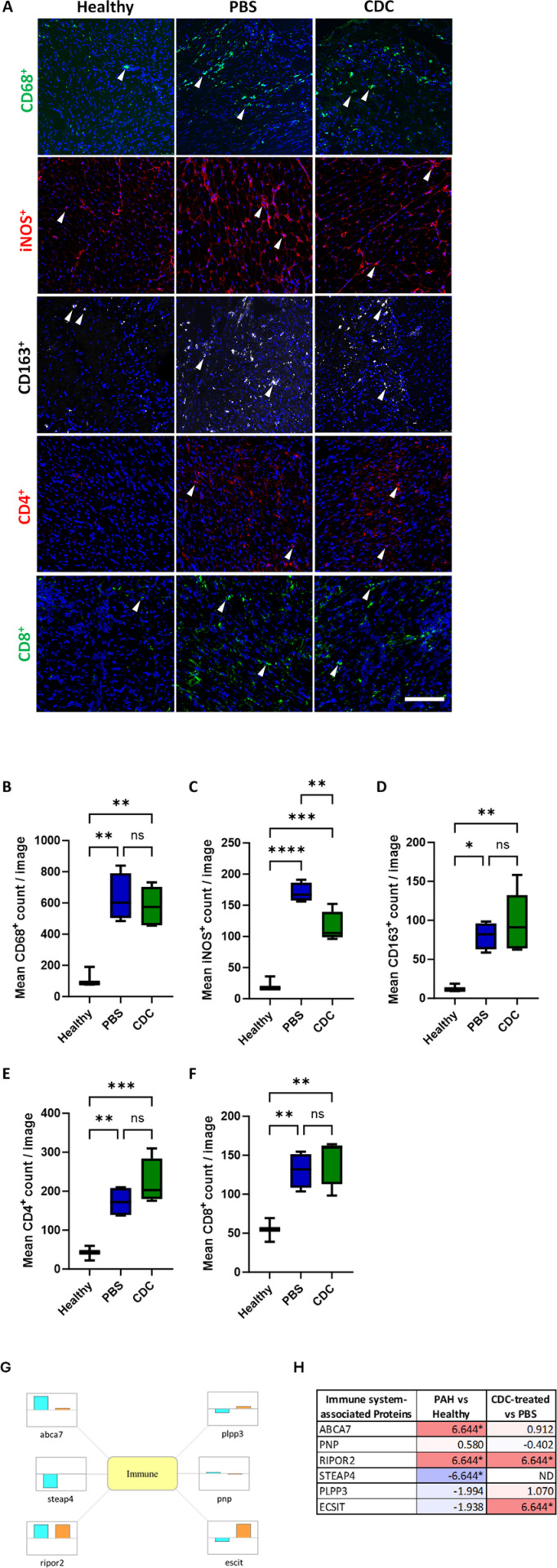
RV muscle capillarity was significantly reduced in the PAH rats while RV muscle fibrosis was increased. IC CDCs significantly increased RV muscle capillarity back to levels noted in healthy rats and reduced RV free wall fibrosis. Further, a significant reduction in iNOS+ (M1) macrophages was also observed within the RV free wall in CDC-treated animals. Proteomic analysis of RV muscle in CDC- or PBS-treated PAH rats showed alterations in protein pathways related to inflammation, fibrosis, autophagy, cell vitality, and angiogenesis. These changes were consistent with putative coordination by a small number of key upstream regulators (MYC, TP53, HNF4A, TGFB1, and KRAS). TAPSE was significantly reduced in PBS-treated animals but was maintained at or above baseline levels in CDC-treated animals.

CONCLUSIONS

CDC therapy can significantly impact the maladaptive milieu of the RV myocardium in advanced PAH, by altering several pathobiological pathways. Such adjunctive therapy, in addition to those employed to reduce pulmonary vascular resistance, would be a great advance in managing RV failure, for which no effective current approved therapies exist.

摘要

背景与目的

在肺动脉高压(PAH)中,右心室(RV)功能是生存的主要决定因素。尽管有当前的治疗方法,但PAH患者的右心室肌肉仍会出现适应性不良变化,最终导致右心室功能障碍和衰竭。本研究的目的是评估冠状动脉内(IC)注射心脏球源细胞(CDC)对减轻右心室肌肉适应性不良病理生物学的影响,并评估右心室功能改善的潜在机制。

方法

两组通过三尖瓣环平面收缩期位移(TAPSE)测量显示右心室功能显著降低的PAH Sugen/低氧大鼠模型,分别接受冠状动脉内注射CDC或PBS安慰剂。采用免疫组织化学方法评估右心室病理生物学变化。此外,运用先进的蛋白质组学技术检测蛋白质信号通路和上游调节因子。

结果

PAH大鼠的右心室肌肉毛细血管密度显著降低,而右心室肌肉纤维化增加。冠状动脉内注射CDC可使右心室肌肉毛细血管密度显著恢复至健康大鼠水平,并减少右心室游离壁纤维化。此外,在接受CDC治疗的动物的右心室游离壁内,诱导型一氧化氮合酶阳性(M1)巨噬细胞也显著减少。对接受CDC或PBS治疗的PAH大鼠的右心室肌肉进行蛋白质组学分析,结果显示与炎症、纤维化、自噬、细胞活力和血管生成相关的蛋白质通路发生了改变。这些变化与少数关键上游调节因子(MYC、TP53、HNF4A、TGFB1和KRAS)的假定协调作用一致。在接受PBS治疗的动物中,TAPSE显著降低,但在接受CDC治疗的动物中,TAPSE维持在基线水平或以上。

结论

CDC治疗可通过改变多种病理生物学途径,显著影响晚期PAH患者右心室心肌的适应性不良环境。除了用于降低肺血管阻力的治疗方法外,这种辅助治疗将是管理右心室衰竭的一大进步,目前尚无有效的获批治疗方法。

https://cdn.ncbi.nlm.nih.gov/pmc/blobs/a1bc/12068596/f1a803de8bf5/pone.0321895.g007.jpg
https://cdn.ncbi.nlm.nih.gov/pmc/blobs/a1bc/12068596/a9b838c22320/pone.0321895.g001.jpg
https://cdn.ncbi.nlm.nih.gov/pmc/blobs/a1bc/12068596/98245bd536cc/pone.0321895.g002.jpg
https://cdn.ncbi.nlm.nih.gov/pmc/blobs/a1bc/12068596/ea75eb3ed35b/pone.0321895.g003.jpg
https://cdn.ncbi.nlm.nih.gov/pmc/blobs/a1bc/12068596/c7bbfc8af3d0/pone.0321895.g004.jpg
https://cdn.ncbi.nlm.nih.gov/pmc/blobs/a1bc/12068596/54b9024e44c6/pone.0321895.g005.jpg
https://cdn.ncbi.nlm.nih.gov/pmc/blobs/a1bc/12068596/74f5f81c6535/pone.0321895.g006.jpg
https://cdn.ncbi.nlm.nih.gov/pmc/blobs/a1bc/12068596/f1a803de8bf5/pone.0321895.g007.jpg

相似文献

1
Impact of cardiosphere-derived cells on the maladapted right ventricular muscle in a rat sugen/hypoxia model of pulmonary hypertension with right ventricular dysfunction.心肌球衍生细胞对大鼠苏金/低氧诱导的伴有右心室功能障碍的肺动脉高压模型中适应不良的右心室心肌的影响。
PLoS One. 2025 May 12;20(5):e0321895. doi: 10.1371/journal.pone.0321895. eCollection 2025.
2
Transcriptomic Analysis of Right Ventricular Remodeling in Two Rat Models of Pulmonary Hypertension: Identification and Validation of Epithelial-to-Mesenchymal Transition in Human Right Ventricular Failure.转录组分析两种肺动脉高压大鼠模型右心室重构:人类右心衰竭中上皮-间充质转化的鉴定和验证。
Circ Heart Fail. 2021 Feb;14(2):e007058. doi: 10.1161/CIRCHEARTFAILURE.120.007058. Epub 2021 Feb 5.
3
Carvedilol improves biventricular fibrosis and function in experimental pulmonary hypertension.卡维地洛可改善实验性肺动脉高压中的双心室纤维化及功能。
J Mol Med (Berl). 2015 Jun;93(6):663-74. doi: 10.1007/s00109-015-1251-9. Epub 2015 Jan 18.
4
Distinct time courses and mechanics of right ventricular hypertrophy and diastolic stiffening in a male rat model of pulmonary arterial hypertension.肺动脉高压雄性大鼠模型右心室肥厚和舒张僵硬的不同时间进程和机制。
Am J Physiol Heart Circ Physiol. 2021 Oct 1;321(4):H702-H715. doi: 10.1152/ajpheart.00046.2021. Epub 2021 Aug 27.
5
Collagen 18A1/Endostatin Expression in the Progression of Right Ventricular Remodeling and Dysfunction in Pulmonary Arterial Hypertension.胶原 18A1/内皮抑素在肺动脉高压右心室重构和功能障碍进展中的表达。
Am J Respir Cell Mol Biol. 2024 Sep;71(3):343-355. doi: 10.1165/rcmb.2024-0039OC.
6
Ultrastructural Changes of the Right Ventricular Myocytes in Pulmonary Arterial Hypertension.肺动脉高压右心室心肌细胞的超微结构变化。
J Am Heart Assoc. 2019 Mar 5;8(5):e011227. doi: 10.1161/JAHA.118.011227.
7
Right Ventricular Stiffening and Anisotropy Alterations in Pulmonary Hypertension: Mechanisms and Relations to Right Heart Failure.肺动脉高压时右心室僵硬度和各向异性改变:机制及其与右心衰竭的关系
J Am Heart Assoc. 2025 Mar 4;14(5):e037126. doi: 10.1161/JAHA.124.037126. Epub 2025 Feb 26.
8
Regional septal hinge-point injury contributes to adverse biventricular interactions in pulmonary hypertension.局部间隔铰链点损伤导致肺动脉高压中的不良双心室相互作用。
Physiol Rep. 2017 Jul;5(14). doi: 10.14814/phy2.13332.
9
Beneficial effects of mesenchymal stem cell delivery via a novel cardiac bioscaffold on right ventricles of pulmonary arterial hypertensive rats.通过新型心脏生物支架递送间充质干细胞对肺动脉高压大鼠右心室的有益作用。
Am J Physiol Heart Circ Physiol. 2019 May 1;316(5):H1005-H1013. doi: 10.1152/ajpheart.00091.2018. Epub 2019 Mar 1.
10
17β-estradiol preserves right ventricular function in rats with pulmonary arterial hypertension: an echocardiographic and histochemical study.17β-雌二醇对肺动脉高压大鼠右心室功能的保护作用:一项超声心动图和组织化学研究
Int J Cardiovasc Imaging. 2019 Mar;35(3):441-450. doi: 10.1007/s10554-018-1468-0. Epub 2018 Oct 22.

本文引用的文献

1
Echocardiographic evaluation of right heart failure which might be associated with DNA damage response in SU5416-hypoxia induced pulmonary hypertension rat model.超声心动图评价 SU5416 低氧诱导肺动脉高压大鼠模型右心衰竭与 DNA 损伤反应的关系。
Respir Res. 2023 Aug 17;24(1):202. doi: 10.1186/s12931-023-02501-7.
2
The evolution of survival of pulmonary arterial hypertension over 15 years.15年间肺动脉高压患者生存率的演变
Pulm Circ. 2022 Oct 1;12(4):e12137. doi: 10.1002/pul2.12137. eCollection 2022 Oct.
3
Altered Cellular Immunity and Differentially Expressed Immune-Related Genes in Patients With Systemic Sclerosis-Associated Pulmonary Arterial Hypertension.
系统性硬化症相关肺动脉高压患者的细胞免疫改变和差异表达的免疫相关基因。
Front Immunol. 2022 May 18;13:868983. doi: 10.3389/fimmu.2022.868983. eCollection 2022.
4
Multi‑omics analysis of right ventricles in rat models of pulmonary arterial hypertension: Consideration of mitochondrial biogenesis by chrysin.多组学分析肺动脉高压大鼠模型的右心室:白杨素对线粒体生物发生的考虑。
Int J Mol Med. 2022 May;49(5). doi: 10.3892/ijmm.2022.5124. Epub 2022 Mar 22.
5
Repeated intravenous cardiosphere-derived cell therapy in late-stage Duchenne muscular dystrophy (HOPE-2): a multicentre, randomised, double-blind, placebo-controlled, phase 2 trial.晚期杜氏肌营养不良症重复静脉注射心脏球衍生细胞治疗(HOPE-2):一项多中心、随机、双盲、安慰剂对照的2期试验。
Lancet. 2022 Mar 12;399(10329):1049-1058. doi: 10.1016/S0140-6736(22)00012-5.
6
Penetrance of Severe Pulmonary Arterial Hypertension in Response to Vascular Endothelial Growth Factor Receptor 2 Blockade in a Genetically Prone Rat Model Is Reduced by Female Sex.在遗传易患大鼠模型中,雌性可降低严重肺动脉高压对血管内皮生长因子受体2阻断的反应性。
J Am Heart Assoc. 2021 Aug 3;10(15):e019488. doi: 10.1161/JAHA.120.019488. Epub 2021 Jul 28.
7
Regulatory T cell activation, proliferation, and reprogramming induced by extracellular vesicles.细胞外囊泡诱导调节性 T 细胞的激活、增殖和重编程。
J Heart Lung Transplant. 2021 Nov;40(11):1387-1395. doi: 10.1016/j.healun.2021.06.005. Epub 2021 Jun 24.
8
The Diverse Roles of TNNI3K in Cardiac Disease and Potential for Treatment.TNNI3K 在心脏疾病中的多种作用及治疗潜力。
Int J Mol Sci. 2021 Jun 15;22(12):6422. doi: 10.3390/ijms22126422.
9
ECSIT is a critical limiting factor for cardiac function.ECSIT 是心脏功能的一个关键限制因素。
JCI Insight. 2021 Jun 22;6(12):142801. doi: 10.1172/jci.insight.142801.
10
Extracellular Vesicles as Therapeutic Agents for Cardiac Fibrosis.细胞外囊泡作为心脏纤维化的治疗剂
Front Physiol. 2020 May 21;11:479. doi: 10.3389/fphys.2020.00479. eCollection 2020.