• 文献检索
  • 文档翻译
  • 深度研究
  • 学术资讯
  • Suppr Zotero 插件Zotero 插件
  • 邀请有礼
  • 套餐&价格
  • 历史记录
应用&插件
Suppr Zotero 插件Zotero 插件浏览器插件Mac 客户端Windows 客户端微信小程序
定价
高级版会员购买积分包购买API积分包
服务
文献检索文档翻译深度研究API 文档MCP 服务
关于我们
关于 Suppr公司介绍联系我们用户协议隐私条款
关注我们

Suppr 超能文献

核心技术专利:CN118964589B侵权必究
粤ICP备2023148730 号-1Suppr @ 2026

文献检索

告别复杂PubMed语法,用中文像聊天一样搜索,搜遍4000万医学文献。AI智能推荐,让科研检索更轻松。

立即免费搜索

文件翻译

保留排版,准确专业,支持PDF/Word/PPT等文件格式,支持 12+语言互译。

免费翻译文档

深度研究

AI帮你快速写综述,25分钟生成高质量综述,智能提取关键信息,辅助科研写作。

立即免费体验

超声心动图评价 SU5416 低氧诱导肺动脉高压大鼠模型右心衰竭与 DNA 损伤反应的关系。

Echocardiographic evaluation of right heart failure which might be associated with DNA damage response in SU5416-hypoxia induced pulmonary hypertension rat model.

机构信息

State Key Laboratory of Respiratory Diseases, National Center for Respiratory Medicine, Guangdong Key Laboratory of Vascular Diseases, National Clinical Research Center for Respiratory Diseases, Guangzhou Institute of Respiratory Health, GMU-GIBH Joint School of Life Sciences, the First Affiliated Hospital of Guangzhou Medical University, Guangzhou Medical University, Guangzhou, 510120, Guangdong, China.

The Jackson Laboratory, Bar Harbor, Maine, 04609, USA.

出版信息

Respir Res. 2023 Aug 17;24(1):202. doi: 10.1186/s12931-023-02501-7.

DOI:10.1186/s12931-023-02501-7
PMID:37592245
原文链接:https://pmc.ncbi.nlm.nih.gov/articles/PMC10433698/
Abstract

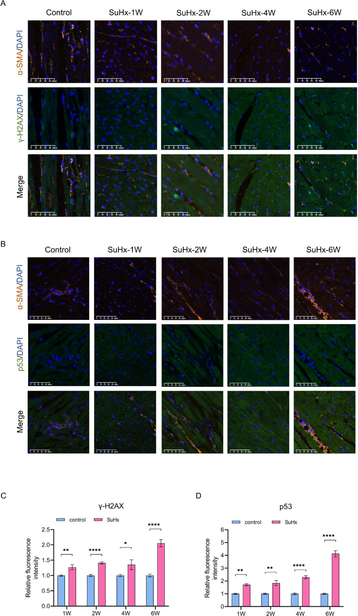
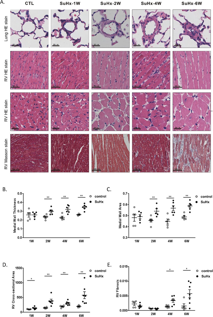
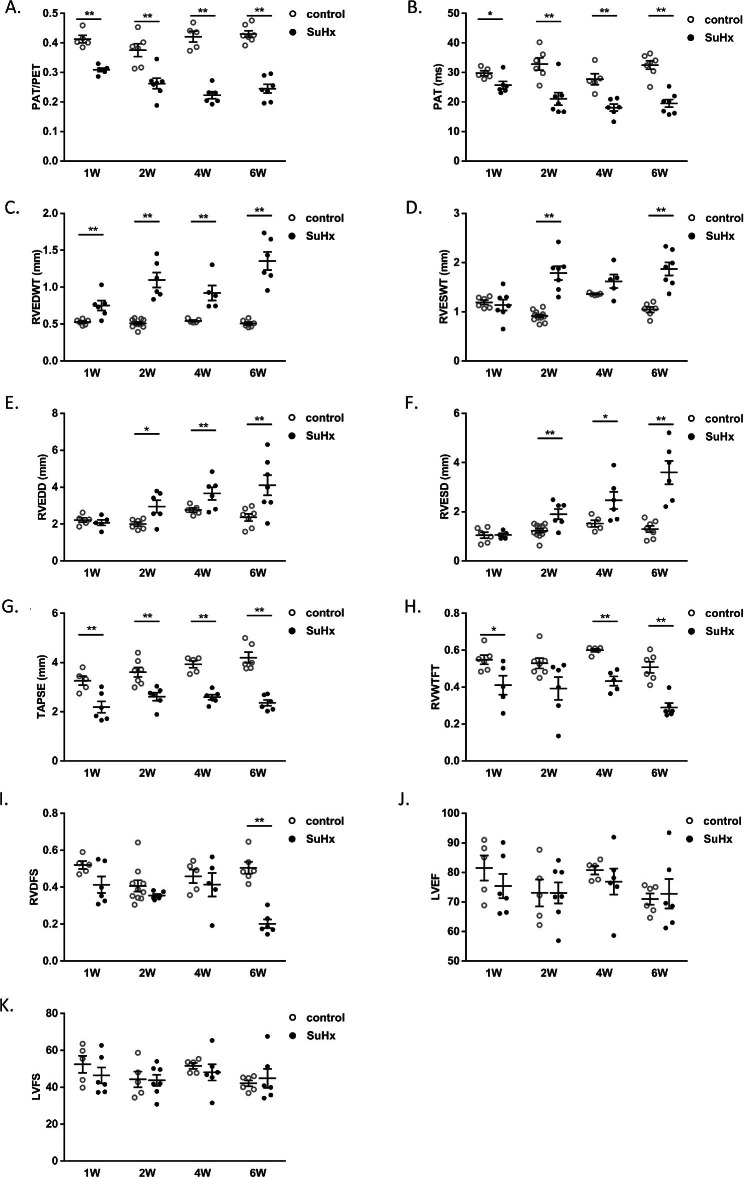
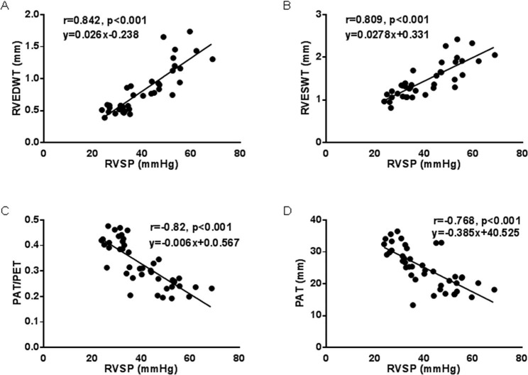
Right heart failure is the leading cause of death in pulmonary hypertension (PH), and echocardiography is a commonly used tool for evaluating the risk hierarchy of PH. However, few studies have explored the dynamic changes in the structural and functional changes of the right heart during the process of PH. Previous studies have found that pulmonary circulation coupling right ventricular adaptation depends on the degree of pressure overload and other factors. In this study, we performed a time-dependent evaluation of right heart functional changes using transthoracic echocardiography in a SU5416 plus hypoxia (SuHx)-induced PH rat model. Rats were examined in 1-, 2-, 4-, and 6-week using right-heart catheterization, cardiac echocardiography, and harvested heart tissue. Our study found that echocardiographic measures of the right ventricle (RV) gradually worsened with the increase of right ventricular systolic pressure, and right heart hypofunction occurred at an earlier stage than pulmonary artery thickening during the development of PH. Furthermore, sarco-endoplasmic reticulum calcium ATPase 2 (SERCA2), a marker of myocardial damage, was highly expressed in week 2 of SuHx-induced PH and had higher levels of expression of γ-H2AX at all timepoints, as well as higher levels of DDR-related proteins p-ATM and p53/p-p53 and p21 in week 4 and week 6. Our study demonstrates that the structure and function of the RV begin to deteriorate with DNA damage and cellular senescence during the early stages of PH development.

摘要

右心衰竭是肺动脉高压(PH)患者死亡的主要原因,超声心动图是评估 PH 风险分层的常用工具。然而,很少有研究探讨 PH 过程中右心结构和功能变化的动态变化。既往研究发现,肺循环耦联右心室适应性取决于压力过载程度等因素。本研究采用 SU5416 加缺氧(SuHx)诱导的 PH 大鼠模型,通过经胸超声心动图对右心功能变化进行时相关评估。通过右心导管、心脏超声和心脏组织采集,在 1、2、4 和 6 周时对大鼠进行检测。我们的研究发现,随着右心室收缩压的升高,右心室超声心动图指标逐渐恶化,在 PH 发展过程中,右心功能障碍发生的时间早于肺动脉壁增厚。此外,肌浆内质网钙 ATP 酶 2(SERCA2)是心肌损伤的标志物,在 SuHx 诱导的 PH 的第 2 周高度表达,在所有时间点 γ-H2AX 的表达水平更高,第 4 周和第 6 周 DDR 相关蛋白 p-ATM 和 p53/p-p53 和 p21 的表达水平也更高。本研究表明,在 PH 早期发展过程中,右心室的结构和功能开始随着 DNA 损伤和细胞衰老而恶化。

https://cdn.ncbi.nlm.nih.gov/pmc/blobs/8f5f/10433698/197df96b6b81/12931_2023_2501_Fig6_HTML.jpg
https://cdn.ncbi.nlm.nih.gov/pmc/blobs/8f5f/10433698/a51e7d871e05/12931_2023_2501_Fig1_HTML.jpg
https://cdn.ncbi.nlm.nih.gov/pmc/blobs/8f5f/10433698/30e4009e8e9e/12931_2023_2501_Fig3_HTML.jpg
https://cdn.ncbi.nlm.nih.gov/pmc/blobs/8f5f/10433698/d430f8884fdf/12931_2023_2501_Fig2_HTML.jpg
https://cdn.ncbi.nlm.nih.gov/pmc/blobs/8f5f/10433698/d0b46914e33a/12931_2023_2501_Fig4_HTML.jpg
https://cdn.ncbi.nlm.nih.gov/pmc/blobs/8f5f/10433698/c4020550a2ba/12931_2023_2501_Fig5_HTML.jpg
https://cdn.ncbi.nlm.nih.gov/pmc/blobs/8f5f/10433698/197df96b6b81/12931_2023_2501_Fig6_HTML.jpg

相似文献

1
Echocardiographic evaluation of right heart failure which might be associated with DNA damage response in SU5416-hypoxia induced pulmonary hypertension rat model.超声心动图评价 SU5416 低氧诱导肺动脉高压大鼠模型右心衰竭与 DNA 损伤反应的关系。
Respir Res. 2023 Aug 17;24(1):202. doi: 10.1186/s12931-023-02501-7.
2
Fischer rats exhibit maladaptive structural and molecular right ventricular remodelling in severe pulmonary hypertension: a genetically prone model for right heart failure.费希尔大鼠在严重肺动脉高压时表现出适应性不良的结构和分子右心室重构:一种易患右心衰竭的遗传性模型。
Cardiovasc Res. 2019 Mar 15;115(4):788-799. doi: 10.1093/cvr/cvy258.
3
Heart Rate Reduction Improves Right Ventricular Function and Fibrosis in Pulmonary Hypertension.心率降低可改善肺动脉高压患者的右心室功能和纤维化。
Am J Respir Cell Mol Biol. 2020 Dec;63(6):843-855. doi: 10.1165/rcmb.2019-0317OC.
4
Sulforaphane prevents right ventricular injury and reduces pulmonary vascular remodeling in pulmonary arterial hypertension.萝卜硫素可预防肺动脉高压右心室损伤并减少肺血管重构。
Am J Physiol Heart Circ Physiol. 2020 Apr 1;318(4):H853-H866. doi: 10.1152/ajpheart.00321.2019. Epub 2020 Feb 28.
5
Isolated heart model demonstrates evidence of contractile and diastolic dysfunction in right ventricles from rats with sugen/hypoxia-induced pulmonary hypertension.离体心脏模型显示,在舒更葡糖/低氧诱导的肺动脉高压大鼠的右心室中存在收缩和舒张功能障碍的证据。
Physiol Rep. 2017 Oct;5(19). doi: 10.14814/phy2.13438. Epub 2017 Oct 16.
6
Down-regulation of the mitochondrial fusion protein Opa1/Mfn2 promotes cardiomyocyte hypertrophy in Su5416/hypoxia-induced pulmonary hypertension rats.下调线粒体融合蛋白 Opa1/Mfn2 促进 Su5416/低氧诱导的肺动脉高压大鼠心肌细胞肥大。
Arch Biochem Biophys. 2023 Oct 1;747:109743. doi: 10.1016/j.abb.2023.109743. Epub 2023 Sep 9.
7
Evaluation of right coronary vascular dysfunction in severe pulmonary hypertensive rats using synchrotron radiation microangiography.应用同步辐射微血管成像技术评价严重肺动脉高压大鼠的右冠状动脉血管功能障碍。
Am J Physiol Heart Circ Physiol. 2021 Mar 1;320(3):H1021-H1036. doi: 10.1152/ajpheart.00327.2020. Epub 2021 Jan 22.
8
Right Ventricular Longitudinal Strain Is Depressed in a Bovine Model of Pulmonary Hypertension.在肺动脉高压的牛模型中右心室纵向应变降低。
Anesth Analg. 2016 May;122(5):1280-6. doi: 10.1213/ANE.0000000000001215.
9
Assessment of right ventricular function in advanced heart failure with nonischemic dilated cardiomyopathy: insights of right ventricular elastance.非缺血性扩张型心肌病所致晚期心力衰竭患者右心室功能的评估:右心室弹性的见解
J Cardiovasc Med (Hagerstown). 2020 Feb;21(2):134-143. doi: 10.2459/JCM.0000000000000921.
10
Three-dimensional echocardiography and 2D-3D speckle-tracking imaging in chronic pulmonary hypertension: diagnostic accuracy in detecting hemodynamic signs of right ventricular (RV) failure.三维超声心动图和二维-三维斑点追踪成像在慢性肺动脉高压中的应用:检测右心室(RV)衰竭血流动力学征象的诊断准确性
J Am Heart Assoc. 2015 Mar 19;4(3):e001584. doi: 10.1161/JAHA.114.001584.

引用本文的文献

1
Impact of cardiosphere-derived cells on the maladapted right ventricular muscle in a rat sugen/hypoxia model of pulmonary hypertension with right ventricular dysfunction.心肌球衍生细胞对大鼠苏金/低氧诱导的伴有右心室功能障碍的肺动脉高压模型中适应不良的右心室心肌的影响。
PLoS One. 2025 May 12;20(5):e0321895. doi: 10.1371/journal.pone.0321895. eCollection 2025.

本文引用的文献

1
Biomarkers of cellular senescence in idiopathic pulmonary fibrosis.特发性肺纤维化中的细胞衰老生物标志物。
Respir Res. 2023 Apr 7;24(1):101. doi: 10.1186/s12931-023-02403-8.
2
Arctiin-encapsulated DSPE-PEG bubble-like nanoparticles inhibit alveolar epithelial type 2 cell senescence to alleviate pulmonary fibrosis the p38/p53/p21 pathway.熊果苷包裹的二硬脂酰磷脂酰乙醇胺-聚乙二醇泡状纳米颗粒通过p38/p53/p21途径抑制肺泡Ⅱ型上皮细胞衰老以减轻肺纤维化。
Front Pharmacol. 2023 Mar 14;14:1141800. doi: 10.3389/fphar.2023.1141800. eCollection 2023.
3
Right Ventricular Architectural Remodeling and Functional Adaptation in Pulmonary Hypertension.
肺动脉高压中的右心室结构重塑和功能适应。
Circ Heart Fail. 2023 Feb;16(2):e009768. doi: 10.1161/CIRCHEARTFAILURE.122.009768. Epub 2023 Feb 7.
4
2022 ESC/ERS Guidelines for the diagnosis and treatment of pulmonary hypertension.2022年欧洲心脏病学会/欧洲呼吸学会肺动脉高压诊断和治疗指南。
Eur Heart J. 2022 Oct 11;43(38):3618-3731. doi: 10.1093/eurheartj/ehac237.
5
The Right Ventricular-Pulmonary Arterial Coupling and Diastolic Function Response to Therapy in Pulmonary Arterial Hypertension.右心室-肺动脉偶联与肺动脉高压治疗后的舒张功能反应。
Chest. 2022 Apr;161(4):1048-1059. doi: 10.1016/j.chest.2021.09.040. Epub 2021 Oct 9.
6
Pulmonary Hypertension in the Context of Heart Failure With Preserved Ejection Fraction.射血分数保留的心力衰竭中的肺动脉高压。
Chest. 2021 Dec;160(6):2232-2246. doi: 10.1016/j.chest.2021.08.039. Epub 2021 Aug 12.
7
DNA Damage and Repair in Pulmonary Arterial Hypertension.DNA 损伤与修复在肺动脉高压中的作用
Genes (Basel). 2020 Oct 19;11(10):1224. doi: 10.3390/genes11101224.
8
Cellular senescence impairs the reversibility of pulmonary arterial hypertension.细胞衰老损害肺动脉高压的可逆性。
Sci Transl Med. 2020 Jul 29;12(554). doi: 10.1126/scitranslmed.aaw4974.
9
Echocardiographic markers of pulmonary hemodynamics and right ventricular hypertrophy in rat models of pulmonary hypertension.肺动脉高压大鼠模型中肺血流动力学和右心室肥厚的超声心动图标志物
Pulm Circ. 2020 May 29;10(2):2045894020910976. doi: 10.1177/2045894020910976. eCollection 2020 Apr-Jun.
10
Cardiomyocyte Senescence and Cellular Communications Within Myocardial Microenvironments.心肌细胞衰老与心肌微环境中的细胞通讯。
Front Endocrinol (Lausanne). 2020 May 21;11:280. doi: 10.3389/fendo.2020.00280. eCollection 2020.