• 文献检索
  • 文档翻译
  • 深度研究
  • 学术资讯
  • Suppr Zotero 插件Zotero 插件
  • 邀请有礼
  • 套餐&价格
  • 历史记录
应用&插件
Suppr Zotero 插件Zotero 插件浏览器插件Mac 客户端Windows 客户端微信小程序
定价
高级版会员购买积分包购买API积分包
服务
文献检索文档翻译深度研究API 文档MCP 服务
关于我们
关于 Suppr公司介绍联系我们用户协议隐私条款
关注我们

Suppr 超能文献

核心技术专利:CN118964589B侵权必究
粤ICP备2023148730 号-1Suppr @ 2026

文献检索

告别复杂PubMed语法,用中文像聊天一样搜索,搜遍4000万医学文献。AI智能推荐,让科研检索更轻松。

立即免费搜索

文件翻译

保留排版,准确专业,支持PDF/Word/PPT等文件格式,支持 12+语言互译。

免费翻译文档

深度研究

AI帮你快速写综述,25分钟生成高质量综述,智能提取关键信息,辅助科研写作。

立即免费体验

一种用于区分肺良性和恶性结节的新型复合模型。

A novel composite model for distinguishing benign and malignant pulmonary nodules.

作者信息

Zhang Lei, Xu Yanhui, Lou Qinqin, Chen Fangfang, Li Fang, Chai Kun, Gao Junshun, Tong Mingjie, Ma Yan, Xia Lilong, Zhao Kaixiang, Gao Junli, Zhu Xinhai

机构信息

Department of Thoracic Surgery, Zhejiang Hospital, Hangzhou, 310013, China.

Hangzhou Cosmos Wisdom Mass Spectrometry Center of Zhejiang University Medical School, Hangzhou, 311200, China.

出版信息

Clin Exp Med. 2025 May 14;25(1):159. doi: 10.1007/s10238-025-01672-5.

DOI:10.1007/s10238-025-01672-5
PMID:40366455
原文链接:https://pmc.ncbi.nlm.nih.gov/articles/PMC12078401/
Abstract

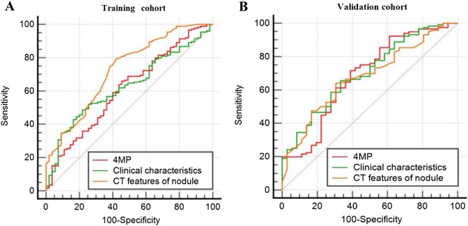
Previous studies have demonstrated that a four-protein marker panel (4MP), consisting of Pro-SFTPB, CA125, Cyfra21-1, and CEA could be used to identify benign and malignant lung nodules. This study aims to improve the 4MP's performance by combining clinical characteristics and low-dose chest computed tomography (LDCT) screening features. This study involved 380 patients with pulmonary nodules, diagnosing 91 benign and 289 early-stage lung cancer via postoperative histopathology. Serum levels of Pro-SFTPB, CA125, Cyfra21-1, and CEA were assessed using an immunofluorescence assay. Clinical features were selected using the LassoCV method. A new diagnostic model was developed using logistic regression, incorporating 4MP, clinical characteristics, and LDCT features. The model's diagnostic performance was compared to the lung cancer biomarker panel (LCBP) nodule risk model, and evaluated through sensitivity, specificity, and the AUC value. The AUC values for distinguishing between benign and malignant pulmonary nodules were 0.612 for the 4MP model. We screened out 7 factors of patient clinical information and CT features of nodules. The composite model (4MP + age + gender + BMI + family history of cancer + nodule size + nodule margin + nodule density) achieved an AUC of 0.808, especially for small nodules (AUC = 0.835 for nodules ≤ 6 mm). Furthermore, within the same validation cohort, the performance of the composite model (AUC = 0.680) surpassed that of the LCBP nodule risk model (AUC = 0.599). The novel composite model accurately diagnoses malignant pulmonary nodules, especially small ones, helping to stratify patients by lung cancer risk.

摘要

先前的研究表明,由表面活性蛋白B前体(Pro-SFTPB)、癌抗原125(CA125)、细胞角蛋白19片段(Cyfra21-1)和癌胚抗原(CEA)组成的四蛋白标志物组合(4MP)可用于鉴别肺结节的良恶性。本研究旨在通过结合临床特征和低剂量胸部计算机断层扫描(LDCT)筛查特征来提高4MP的性能。本研究纳入了380例肺结节患者,通过术后组织病理学诊断出91例良性结节和289例早期肺癌。采用免疫荧光法检测血清中Pro-SFTPB、CA125、Cyfra21-1和CEA的水平。使用套索交叉验证(LassoCV)方法选择临床特征。采用逻辑回归建立了一个新的诊断模型,纳入了4MP、临床特征和LDCT特征。将该模型的诊断性能与肺癌生物标志物组合(LCBP)结节风险模型进行比较,并通过敏感性、特异性和曲线下面积(AUC)值进行评估。4MP模型区分良性和恶性肺结节的AUC值为0.612。我们筛选出了7个患者临床信息和结节CT特征的因素。复合模型(4MP + 年龄 + 性别 + 体重指数 + 癌症家族史 + 结节大小 + 结节边缘 + 结节密度)的AUC为0.808,尤其是对于小结节(直径≤6 mm的结节AUC = 0.835)。此外,在同一验证队列中,复合模型(AUC = 0.680)的性能超过了LCBP结节风险模型(AUC = 0.599)。这种新型复合模型能够准确诊断恶性肺结节,尤其是小结节,有助于根据肺癌风险对患者进行分层。

https://cdn.ncbi.nlm.nih.gov/pmc/blobs/122b/12078401/6ca416ae63f1/10238_2025_1672_Fig6_HTML.jpg
https://cdn.ncbi.nlm.nih.gov/pmc/blobs/122b/12078401/d58a54522740/10238_2025_1672_Fig1_HTML.jpg
https://cdn.ncbi.nlm.nih.gov/pmc/blobs/122b/12078401/c91641d92334/10238_2025_1672_Fig2_HTML.jpg
https://cdn.ncbi.nlm.nih.gov/pmc/blobs/122b/12078401/83d34a69ac1f/10238_2025_1672_Fig3_HTML.jpg
https://cdn.ncbi.nlm.nih.gov/pmc/blobs/122b/12078401/4f37165229dd/10238_2025_1672_Fig4_HTML.jpg
https://cdn.ncbi.nlm.nih.gov/pmc/blobs/122b/12078401/c766ffd78774/10238_2025_1672_Fig5_HTML.jpg
https://cdn.ncbi.nlm.nih.gov/pmc/blobs/122b/12078401/6ca416ae63f1/10238_2025_1672_Fig6_HTML.jpg

相似文献

1
A novel composite model for distinguishing benign and malignant pulmonary nodules.一种用于区分肺良性和恶性结节的新型复合模型。
Clin Exp Med. 2025 May 14;25(1):159. doi: 10.1007/s10238-025-01672-5.
2
Combining serum miRNAs, CEA, and CYFRA21-1 with imaging and clinical features to distinguish benign and malignant pulmonary nodules: a pilot study : Xianfeng Li et al.: Combining biomarker, imaging, and clinical features to distinguish pulmonary nodules.联合血清微小RNA、癌胚抗原和细胞角蛋白19片段与影像学及临床特征鉴别肺结节的良恶性:一项初步研究:李先锋等人:联合生物标志物、影像学和临床特征鉴别肺结节
World J Surg Oncol. 2017 May 25;15(1):107. doi: 10.1186/s12957-017-1171-y.
3
Diagnostic value of conventional tumor markers in young patients with pulmonary nodules.常规肿瘤标志物对肺部结节年轻患者的诊断价值。
J Clin Lab Anal. 2021 Sep;35(9):e23912. doi: 10.1002/jcla.23912. Epub 2021 Jul 23.
4
[Value of Combined Detection of Cytokines and Tumor Markers in the 
Differential Diagnosis of Benign and Malignant Solitary Pulmonary Nodules].[细胞因子与肿瘤标志物联合检测在孤立性肺结节良恶性鉴别诊断中的价值]
Zhongguo Fei Ai Za Zhi. 2021 Jun 20;24(6):426-433. doi: 10.3779/j.issn.1009-3419.2021.102.20.
5
[Diagnostic value of serum tumor markers in differentiating malignant from benign solitary pulmonary nodules].[血清肿瘤标志物在鉴别孤立性肺结节良恶性中的诊断价值]
Beijing Da Xue Xue Bao Yi Xue Ban. 2014 Oct 18;46(5):707-10.
6
Enhancing the differential diagnosis of small pulmonary nodules: a comprehensive model integrating plasma methylation, protein biomarkers, and LDCT imaging features.提高肺部小结节的鉴别诊断能力:综合血浆甲基化、蛋白生物标志物和 LDCT 成像特征的模型。
J Transl Med. 2024 Oct 31;22(1):984. doi: 10.1186/s12967-024-05723-5.
7
A combined model of circulating tumor DNA methylated SHOX2/SCT/HOXA7 and clinical features facilitates the discrimination of malignant from benign pulmonary nodules.循环肿瘤DNA甲基化的SHOX2/SCT/HOXA7与临床特征的联合模型有助于鉴别恶性与良性肺结节。
Lung Cancer. 2025 Jan;199:108064. doi: 10.1016/j.lungcan.2024.108064. Epub 2024 Dec 16.
8
Preoperative diagnosis of malignant pulmonary nodules in lung cancer screening with a radiomics nomogram.肺癌筛查中基于放射组学列线图的恶性肺结节术前诊断。
Cancer Commun (Lond). 2020 Jan;40(1):16-24. doi: 10.1002/cac2.12002. Epub 2020 Mar 3.
9
A prediction model based on DNA methylation biomarkers and radiological characteristics for identifying malignant from benign pulmonary nodules.基于 DNA 甲基化生物标志物和放射学特征的预测模型,用于鉴别良恶性肺结节。
BMC Cancer. 2021 Mar 10;21(1):263. doi: 10.1186/s12885-021-08002-4.
10
A nomogram combining CT-based radiomic features with clinical features for the differentiation of benign and malignant cystic pulmonary nodules.一种结合 CT 影像组学特征和临床特征的列线图模型,用于区分良恶性肺囊性结节。
J Cardiothorac Surg. 2024 Jun 27;19(1):392. doi: 10.1186/s13019-024-02936-z.

本文引用的文献

1
Accurate Diagnosis of High-Risk Pulmonary Nodules Using a Non-Invasive Epigenetic Biomarker Test.使用非侵入性表观遗传生物标志物检测准确诊断高危肺结节
Cancers (Basel). 2025 Mar 7;17(6):916. doi: 10.3390/cancers17060916.
2
Construction and validation of a risk stratification model based on Lung-RADS v2022 and CT features for predicting the invasive pure ground-glass pulmonary nodules in China.基于Lung-RADS v2022和CT特征构建及验证用于预测中国纯磨玻璃浸润性肺结节的风险分层模型
Insights Imaging. 2025 Mar 23;16(1):68. doi: 10.1186/s13244-025-01937-3.
3
CT radiomics based model for differentiating malignant and benign small (≤20mm) solid pulmonary nodules.
基于CT影像组学的模型用于鉴别直径≤20mm的肺部实性小结节的良恶性
Front Oncol. 2025 Feb 13;15:1502932. doi: 10.3389/fonc.2025.1502932. eCollection 2025.
4
MAEMC-NET: a hybrid self-supervised learning method for predicting the malignancy of solitary pulmonary nodules from CT images.MAEMC-NET:一种用于从CT图像预测孤立性肺结节恶性程度的混合自监督学习方法。
Front Med (Lausanne). 2025 Feb 12;12:1507258. doi: 10.3389/fmed.2025.1507258. eCollection 2025.
5
How I Do It: Management of Pleural-attached Pulmonary Nodules in Low-Dose CT Screening for Lung Cancer.我的做法:低剂量CT肺癌筛查中胸膜附着型肺结节的管理
Radiology. 2025 Jan;314(1):e240091. doi: 10.1148/radiol.240091.
6
Diameter thresholds for pure ground-glass pulmonary nodules at low-dose CT screening: Chinese experience.低剂量CT筛查中纯磨玻璃肺结节的直径阈值:中国经验
Thorax. 2025 Jan 17;80(2):76-85. doi: 10.1136/thorax-2024-221642.
7
Dual biomarkers CT-based deep learning model incorporating intrathoracic fat for discriminating benign and malignant pulmonary nodules in multi-center cohorts.基于CT的深度学习模型结合胸腔内脂肪用于多中心队列中鉴别良性和恶性肺结节的双重生物标志物。
Phys Med. 2025 Jan;129:104877. doi: 10.1016/j.ejmp.2024.104877. Epub 2024 Dec 16.
8
Guidelines for the management of pulmonary nodules detected by low-dose CT lung cancer screening 6th edition: compiled by the Japanese Society of CT Screening.低剂量CT肺癌筛查发现的肺结节管理指南第6版:由日本CT筛查学会编制。
Jpn J Radiol. 2025 Mar;43(3):333-346. doi: 10.1007/s11604-024-01695-0. Epub 2024 Dec 5.
9
Lung Nodule Management in Low-Dose CT Screening for Lung Cancer: Lessons from the NELSON Trial.低剂量 CT 筛查肺癌中的肺结节管理:来自 NELSON 试验的经验。
Radiology. 2024 Oct;313(1):e240535. doi: 10.1148/radiol.240535.
10
Biomarker trajectory for earlier detection of lung cancer.生物标志物轨迹用于早期肺癌检测。
EBioMedicine. 2024 Oct;108:105377. doi: 10.1016/j.ebiom.2024.105377. Epub 2024 Sep 30.