• 文献检索
  • 文档翻译
  • 深度研究
  • 学术资讯
  • Suppr Zotero 插件Zotero 插件
  • 邀请有礼
  • 套餐&价格
  • 历史记录
应用&插件
Suppr Zotero 插件Zotero 插件浏览器插件Mac 客户端Windows 客户端微信小程序
定价
高级版会员购买积分包购买API积分包
服务
文献检索文档翻译深度研究API 文档MCP 服务
关于我们
关于 Suppr公司介绍联系我们用户协议隐私条款
关注我们

Suppr 超能文献

核心技术专利:CN118964589B侵权必究
粤ICP备2023148730 号-1Suppr @ 2026

文献检索

告别复杂PubMed语法,用中文像聊天一样搜索,搜遍4000万医学文献。AI智能推荐,让科研检索更轻松。

立即免费搜索

文件翻译

保留排版,准确专业,支持PDF/Word/PPT等文件格式,支持 12+语言互译。

免费翻译文档

深度研究

AI帮你快速写综述,25分钟生成高质量综述,智能提取关键信息,辅助科研写作。

立即免费体验

PET 影像组学特征在新诊断口腔鳞状细胞癌诊断及复发预测中的价值

Value of PET radiomic features for diagnosis and reccurence prediction of newly diagnosed oral squamous cell carcinoma.

作者信息

Pfaehler Elisabeth, Schindele Andreas, Dierks Alexander, Busse Cornelius, Brumberg Joachim, Kübler Alexander C, Buck Andreas K, Linz Christian, Lapa Constantin, Brands Roman C, Kertels Olivia

机构信息

Nuclear Medicine, Faculty of Medicine, University of Augsburg, Augsburg, Germany.

Department of Oral and Maxillofacial Plastic Surgery, University Hospital of Würzburg, Würzburg, Germany.

出版信息

Sci Rep. 2025 May 20;15(1):17475. doi: 10.1038/s41598-025-02305-3.

DOI:10.1038/s41598-025-02305-3
PMID:40394092
原文链接:https://pmc.ncbi.nlm.nih.gov/articles/PMC12092716/
Abstract

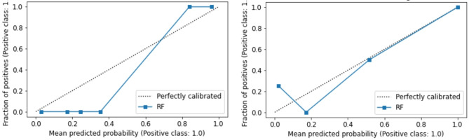
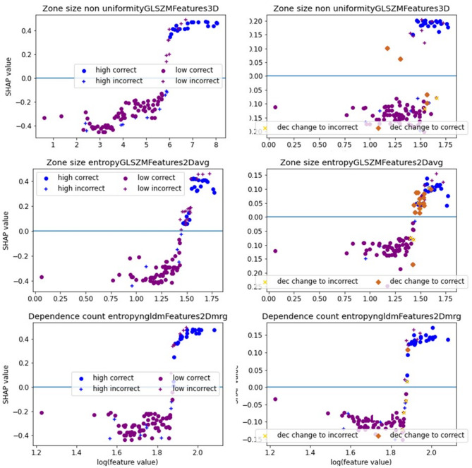
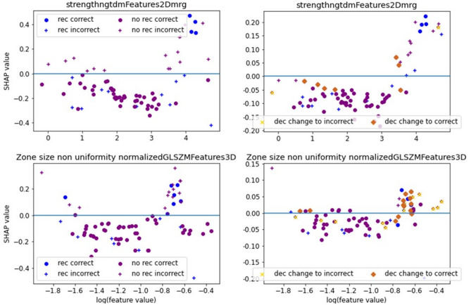
Oral Squamous Cell Carcinoma (OSCC) represents more than 90% of oral cancers. The usefulness of radiomic features extracted from PET images of OSCC patients to predict tumor characteristics such as primary tumor stage (T-stage), or tumor grade has not been investigated yet. In this prospective study, 112 patients with newly diagnosed, treatment-naïve OSCC were included. Tumor segmentation was performed using three strategies, the majority vote of these segmentations was used to calculate 445 radiomic features. Features instable over segmentation methods and features highly correlated with volume, SUV, and SUV were eliminated. A Random Forest classifier was trained to predict T-stage, tumor grade, lymph node involvement, and tumor recurrence. Stratified 10-fold cross-validation was performed. Evaluation metrics such as accuracy and area under the curve (AUC) were reported. SHAP dependence plots were generated to understand classifier decisions. The classifier reached a mean cross-validation AUC of 0.83 for predicting T-stage, an AUC of 0.56 for the grading of the primary tumor, a mean AUC of 0.64 for lymph node involvement, and a mean AUC of 0.63 for recurrence. In patients with newly-diagnosed OSCC, radiomics might have some potential to predict T-stage. These results need to be validated in a larger patient cohort.

摘要

口腔鳞状细胞癌(OSCC)占口腔癌的90%以上。从OSCC患者的PET图像中提取的放射组学特征用于预测肿瘤特征(如原发肿瘤分期(T分期)或肿瘤分级)的有效性尚未得到研究。在这项前瞻性研究中,纳入了112例新诊断的、未接受过治疗的OSCC患者。使用三种策略进行肿瘤分割,这些分割的多数投票结果用于计算445个放射组学特征。消除了在分割方法上不稳定的特征以及与体积、SUV和SUV高度相关的特征。训练了一个随机森林分类器来预测T分期、肿瘤分级、淋巴结受累情况和肿瘤复发情况。进行了分层10折交叉验证。报告了诸如准确率和曲线下面积(AUC)等评估指标。生成了SHAP依赖图以了解分类器的决策。该分类器预测T分期的平均交叉验证AUC为0.83,原发肿瘤分级的AUC为0.56,淋巴结受累的平均AUC为0.64,复发的平均AUC为0.63。在新诊断的OSCC患者中,放射组学可能具有预测T分期的一些潜力。这些结果需要在更大的患者队列中进行验证。

https://cdn.ncbi.nlm.nih.gov/pmc/blobs/fc0b/12092716/028cc98973e6/41598_2025_2305_Fig8_HTML.jpg
https://cdn.ncbi.nlm.nih.gov/pmc/blobs/fc0b/12092716/7324a697848e/41598_2025_2305_Fig1_HTML.jpg
https://cdn.ncbi.nlm.nih.gov/pmc/blobs/fc0b/12092716/7778df55d4f0/41598_2025_2305_Fig2_HTML.jpg
https://cdn.ncbi.nlm.nih.gov/pmc/blobs/fc0b/12092716/651de551fb44/41598_2025_2305_Fig3_HTML.jpg
https://cdn.ncbi.nlm.nih.gov/pmc/blobs/fc0b/12092716/a22a7ca01df1/41598_2025_2305_Fig4_HTML.jpg
https://cdn.ncbi.nlm.nih.gov/pmc/blobs/fc0b/12092716/e2223bdc2e53/41598_2025_2305_Fig5_HTML.jpg
https://cdn.ncbi.nlm.nih.gov/pmc/blobs/fc0b/12092716/e65f52d0fa66/41598_2025_2305_Fig6_HTML.jpg
https://cdn.ncbi.nlm.nih.gov/pmc/blobs/fc0b/12092716/916ec80d41fb/41598_2025_2305_Fig7_HTML.jpg
https://cdn.ncbi.nlm.nih.gov/pmc/blobs/fc0b/12092716/028cc98973e6/41598_2025_2305_Fig8_HTML.jpg

相似文献

1
Value of PET radiomic features for diagnosis and reccurence prediction of newly diagnosed oral squamous cell carcinoma.PET 影像组学特征在新诊断口腔鳞状细胞癌诊断及复发预测中的价值
Sci Rep. 2025 May 20;15(1):17475. doi: 10.1038/s41598-025-02305-3.
2
MRI-based deep learning and radiomics for prediction of occult cervical lymph node metastasis and prognosis in early-stage oral and oropharyngeal squamous cell carcinoma: a diagnostic study.基于 MRI 的深度学习和放射组学预测早期口腔和口咽鳞状细胞癌隐匿性颈部淋巴结转移和预后的诊断研究。
Int J Surg. 2024 Aug 1;110(8):4648-4659. doi: 10.1097/JS9.0000000000001578.
3
Development and validation of an explainable machine learning model for predicting occult lymph node metastasis in early-stage oral tongue squamous cell carcinoma: A multi-center study.用于预测早期口腔舌鳞状细胞癌隐匿性淋巴结转移的可解释机器学习模型的开发与验证:一项多中心研究
Int J Surg. 2025 Aug 1;111(8):5022-5035. doi: 10.1097/JS9.0000000000002641. Epub 2025 Jun 5.
4
Radiomics Nomogram Based on Optimal Volume of Interest Derived from High-Resolution CT for Preoperative Prediction of IASLC Grading in Clinical IA Lung Adenocarcinomas: A Multi-Center, Large-Population Study.基于高分辨率 CT 最优感兴趣区体积的放射组学列线图预测临床 IA 期肺腺癌 IASLC 分级:多中心大样本研究。
Technol Cancer Res Treat. 2024 Jan-Dec;23:15330338241300734. doi: 10.1177/15330338241300734.
5
Comparison of Two Modern Survival Prediction Tools, SORG-MLA and METSSS, in Patients With Symptomatic Long-bone Metastases Who Underwent Local Treatment With Surgery Followed by Radiotherapy and With Radiotherapy Alone.两种现代生存预测工具 SORG-MLA 和 METSSS 在接受手术联合放疗和单纯放疗治疗有症状长骨转移患者中的比较。
Clin Orthop Relat Res. 2024 Dec 1;482(12):2193-2208. doi: 10.1097/CORR.0000000000003185. Epub 2024 Jul 23.
6
Radiomics based on dual-energy CT for noninvasive prediction of cervical lymph node metastases in patients with nasopharyngeal carcinoma.基于双能CT的影像组学对鼻咽癌患者颈部淋巴结转移的无创预测
Radiography (Lond). 2025 Jul;31(4):102989. doi: 10.1016/j.radi.2025.102989. Epub 2025 May 26.
7
Application value of dual-sequence MRI based nomogram of radiomics and morphologic features in predicting tumor differentiation degree and lymph node metastasis of Oral squamous cell carcinoma.基于影像组学和形态学特征的双序列MRI列线图在预测口腔鳞状细胞癌肿瘤分化程度和淋巴结转移中的应用价值
Front Oncol. 2025 Jul 15;15:1588358. doi: 10.3389/fonc.2025.1588358. eCollection 2025.
8
Stable and discriminating OCT-derived radiomics features for predicting anti-VEGF treatment response in diabetic macular edema.用于预测糖尿病性黄斑水肿抗VEGF治疗反应的稳定且有鉴别力的光学相干断层扫描衍生的放射组学特征。
Med Phys. 2025 May;52(5):2762-2772. doi: 10.1002/mp.17695. Epub 2025 Mar 14.
9
Radiomics-based prediction of T cell-inflamed gene expression profile and prognosis in head and neck squamous cell carcinoma.基于影像组学对头颈部鳞状细胞癌中T细胞炎症基因表达谱及预后的预测
Med Phys. 2025 Aug;52(8):e18028. doi: 10.1002/mp.18028.
10
Bioinformatics identification and validation of m6A/m1A/m5C/m7G/ac4 C-modified genes in oral squamous cell carcinoma.口腔鳞状细胞癌中m6A/m1A/m5C/m7G/ac4C修饰基因的生物信息学鉴定与验证
BMC Cancer. 2025 Jul 1;25(1):1055. doi: 10.1186/s12885-025-14216-7.

本文引用的文献

1
Radiomic features of dynamic contrast-enhanced MRI can predict Ki-67 status in head and neck squamous cell carcinoma.动态对比增强磁共振成像的影像组学特征可预测头颈部鳞状细胞癌中的Ki-67状态。
Magn Reson Imaging. 2025 Feb;116:110276. doi: 10.1016/j.mri.2024.110276. Epub 2024 Nov 19.
2
FDG PET-CT for the Detection of Occult Nodal Metastases in Head and Neck Cancer: A Systematic Review and Meta-Analysis.氟代脱氧葡萄糖正电子发射断层扫描-计算机断层扫描用于检测头颈癌隐匿性淋巴结转移:一项系统评价和荟萃分析
Cancers (Basel). 2024 Aug 24;16(17):2954. doi: 10.3390/cancers16172954.
3
Prediction of Histological Grade of Oral Squamous Cell Carcinoma Using Machine Learning Models Applied to F-FDG-PET Radiomics.
使用应用于F-FDG-PET放射组学的机器学习模型预测口腔鳞状细胞癌的组织学分级
Biomedicines. 2024 Jun 25;12(7):1411. doi: 10.3390/biomedicines12071411.
4
Role of F-FDG PET/CT in Head and Neck Squamous Cell Carcinoma: Current Evidence and Innovative Applications.F-FDG PET/CT在头颈部鳞状细胞癌中的作用:当前证据与创新应用
Cancers (Basel). 2024 May 16;16(10):1905. doi: 10.3390/cancers16101905.
5
Is PET Radiomics Useful to Predict Pathologic Tumor Response and Prognosis in Locally Advanced Cervical Cancer?PET 放射组学在预测局部晚期宫颈癌的病理肿瘤反应和预后方面是否有用?
J Nucl Med. 2024 Jun 3;65(6):962-970. doi: 10.2967/jnumed.123.267044.
6
Prognostic value of F-FDG PET radiomics and sarcopenia in patients with oral squamous cell carcinoma.18F-FDG PET 影像组学与骨骼肌减少症对口腔鳞状细胞癌患者预后的预测价值。
Med Phys. 2024 Jul;51(7):4907-4921. doi: 10.1002/mp.16949. Epub 2024 Jan 22.
7
Identification of CT-based non-invasive radiomic biomarkers for overall survival prediction in oral cavity squamous cell carcinoma.基于 CT 的无创放射组学生物标志物用于预测口腔鳞状细胞癌的总生存期。
Sci Rep. 2023 Dec 8;13(1):21774. doi: 10.1038/s41598-023-48048-x.
8
Optimizing Clinical Decision Making with Decision Curve Analysis: Insights for Clinical Investigators.运用决策曲线分析优化临床决策:给临床研究者的见解
Healthcare (Basel). 2023 Aug 10;11(16):2244. doi: 10.3390/healthcare11162244.
9
The prognostic value of radiomic features from pre- and post-treatment F-FDG PET imaging in patients with nasopharyngeal carcinoma.鼻咽癌患者治疗前后 F-FDG PET 影像的放射组学特征的预后价值。
Sci Rep. 2023 May 25;13(1):8462. doi: 10.1038/s41598-023-35582-x.
10
Tumor Recurrence and Follow-Up Intervals in Oral Squamous Cell Carcinoma.口腔鳞状细胞癌的肿瘤复发与随访间隔
J Clin Med. 2022 Nov 29;11(23):7061. doi: 10.3390/jcm11237061.