Jabbour Richard J, Behradfar Elham, Debney Michael, Nygren Anders, Hartley Adam, Efimov Igor, Hocini Mélèze, Peters Nicholas S, Ng Fu Siong, Vigmond Edward J
National Heart and Lung Institute, Imperial College London, London, United Kingdom.
Department of Biomedical Engineering, University of Calgary, Calgary, AB, Canada.
Front Physiol. 2025 May 6;16:1540400. doi: 10.3389/fphys.2025.1540400. eCollection 2025.
The Purkinje network is essential for normal electrical impulse propagation in the heart but has also been implicated in ventricular arrhythmias. Previous experimental work has suggested that not all Purkinje-myocardial junctions (PMJs) are active at rest due to source-sink mismatch at the PMJs.
We hypothesized that pathological conditions that cause gap junction uncoupling (e.g., acute ischaemia), would increase the number of active PMJs, leading to more complex activation patterns.
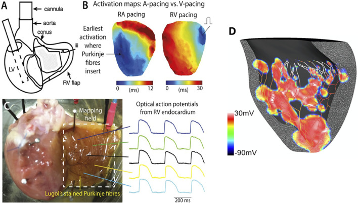
We investigated this using a whole-heart intact Purkinje system preparation that allowed direct high-resolution endocardial mapping to interrogate PMJ function. Twelve (7 control, five rotigaptide) Langendorff-perfused hearts from New Zealand white rabbits were subjected to an ischaemia-reperfusion protocol and optically mapped. Computational modelling was performed to determine the effects of gap junction coupling on PMJ function, and on the complexity of endocardial activation.
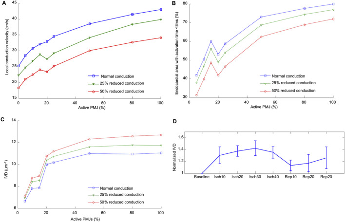
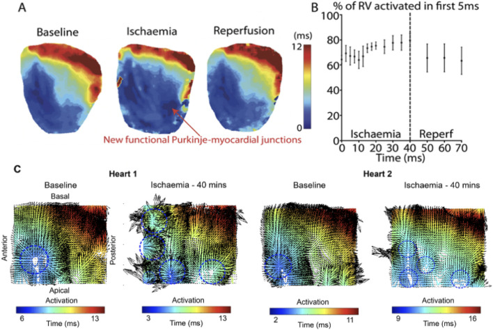
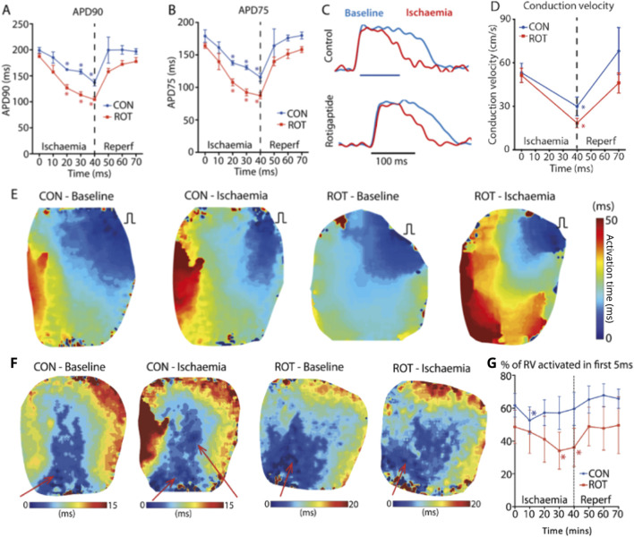
During ischaemia, the percentage of right ventricle area activated within the first 5 ms decreased from baseline 62% ± 7% to 52% ± 8% during early ischaemia (p = 0.04), consistent with slowing of conduction. This was followed by a paradoxical increase in late-ischaemia (60% ± 8%) due to extra regions of early activation. Gap junction enhancement with rotigaptide during ischaemia abolished the aforementioned pattern. Parallel computational experiments replicated experimental findings only when the number of functional PMJs was increased during ischaemia. With more active PMJs, there were more breakthrough sites with increased complexity of activation, as also measured in biological preparations.
Normally-quiescent PMJs can become active in the context of gap junction uncoupling during acute ischaemia. Pharmacological gap junction modulation may alter propagation patterns across PMJs and may be used as a therapeutic strategy for Purkinje system associated arrhythmias.
浦肯野网络对于心脏正常电冲动的传播至关重要,但也与室性心律失常有关。先前的实验研究表明,由于浦肯野-心肌连接点(PMJ)处的源-汇不匹配,并非所有的浦肯野-心肌连接点在静息时都是活跃的。
我们假设导致缝隙连接解偶联的病理状况(如急性缺血)会增加活跃PMJ的数量,从而导致更复杂的激活模式。
我们使用全心完整浦肯野系统制备方法对此进行研究,该方法允许进行直接的高分辨率心内膜标测以探究PMJ功能。对12只(7只对照,5只罗替加肽处理)来自新西兰白兔的Langendorff灌注心脏进行缺血-再灌注方案处理并进行光学标测。进行计算建模以确定缝隙连接耦联对PMJ功能以及心内膜激活复杂性的影响。
在缺血期间,右心室在前5毫秒内被激活的面积百分比从基线时的62%±7%在缺血早期降至52%±8%(p = 0.04),这与传导减慢一致。随后在缺血后期出现反常增加(60%±8%),这是由于早期激活的额外区域所致。缺血期间使用罗替加肽增强缝隙连接消除了上述模式。平行的计算实验仅在缺血期间功能性PMJ数量增加时才复制了实验结果。随着更多的PMJ活跃,有更多的突破位点,激活复杂性增加,这在生物制剂中也得到了测量。
在急性缺血期间,正常情况下静止的PMJ在缝隙连接解偶联的情况下可能会变得活跃。药理学上对缝隙连接的调节可能会改变跨PMJ的传播模式,并可作为治疗与浦肯野系统相关心律失常的一种治疗策略。