Wang Yi-Fan, Zheng Dan, Zhang Ying, Li Xiao-Fen, Xia Ming, Tang Hai-Ming, Huang Chun-Hua, Li Mao-Juan, Lou Di-Dong
Judicial Appraisal Center, Guizhou University of Traditional Chinese Medicine, Guiyang, Guizhou, China.
Key Laboratory of Forensic Toxicology of Herbal Medicines, Guizhou Education Department, Guiyang, Guizhou, China.
Front Pharmacol. 2025 May 13;16:1571960. doi: 10.3389/fphar.2025.1571960. eCollection 2025.
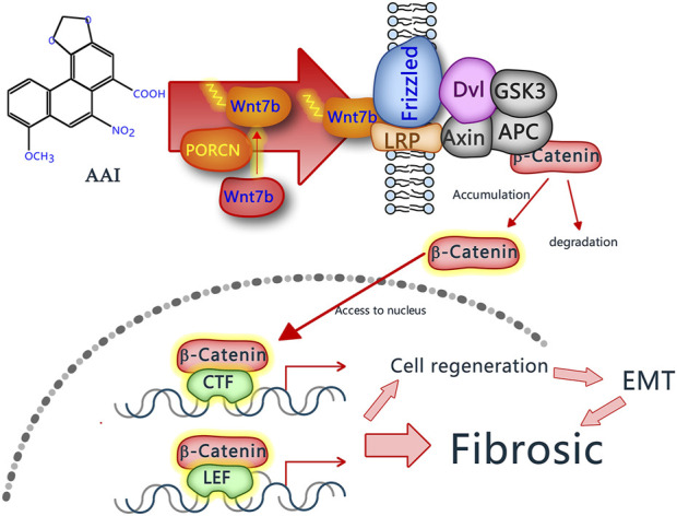
This study investigates the pathological progressions in kidneys affected by aristolochic acid nephropathy (AAN) and explores the molecular mechanisms underlying the fibrotic process, specifically focusing on the Wnt7b/β-catenin signaling pathway.
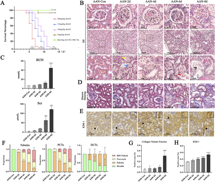
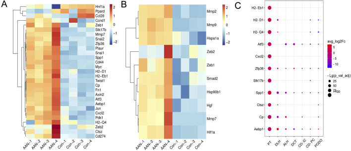
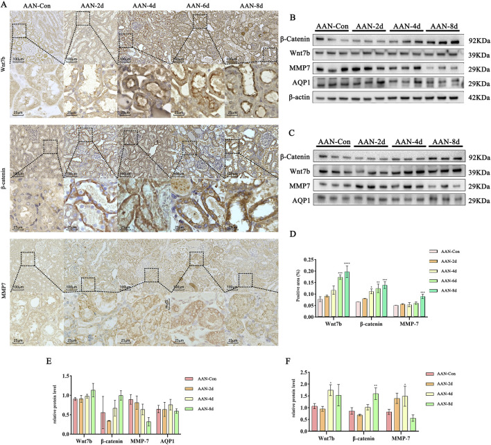
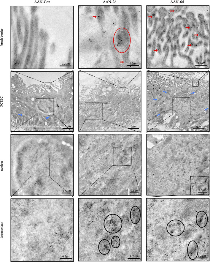
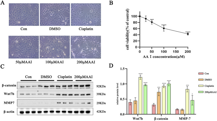
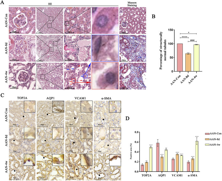
Both mice and human kidney-2 (HK-2) cells were treated with aristolochic acid I (AAI). In mice, we monitored blood urea nitrogen (BUN), serum creatinine (Scr), kidney injury molecule-1 (KIM-1), pathological modifications of renal tubular epithelial cells (RTECs), and fibrosis degrees during acute/chronic disease phases. Wnt7b/β-catenin expression was evaluated through transcriptome analysis and laboratory assays (immunohistochemistry, Western blotting, immunoelectron microscopy) in acute AAN and cultured cells. Concurrent assays measured representative proteins: Aquaporin 1 (AQP1), Topoisomerase IIα (TOP2A), Vascular Cell Adhesion Molecule-1 (VCAM-1), and α-smooth muscle actin (α-SMA) in chronic AAN RTECs.
AAI increased Scr, BUN, and KIM-1 levels by causing RTEC necrotic shedding in acute stages and promoted renal interstitial fibrosis chronically. Elevated Wnt7b pathway proteins enhanced damaged RTEC regeneration, with regenerated cells expressing mesenchymal proteins VCAM-1 and α-SMA.
The Wnt7b/β-catenin signaling pathway connects acute tubule damage to fibrosis, explaining AAN's pathological continuum. These findings clarify how acute injury progresses to chronic fibrosis in AAN.
本研究调查了受马兜铃酸肾病(AAN)影响的肾脏的病理进展,并探索纤维化过程背后的分子机制,特别关注Wnt7b/β-连环蛋白信号通路。
用马兜铃酸I(AAI)处理小鼠和人肾2(HK-2)细胞。在小鼠中,我们监测了急性/慢性疾病阶段的血尿素氮(BUN)、血清肌酐(Scr)、肾损伤分子-1(KIM-1)、肾小管上皮细胞(RTECs)的病理改变以及纤维化程度。通过转录组分析和实验室检测(免疫组织化学、蛋白质印迹法、免疫电子显微镜)评估急性AAN和培养细胞中Wnt7b/β-连环蛋白的表达。同时检测慢性AAN的RTECs中的代表性蛋白质:水通道蛋白1(AQP1)、拓扑异构酶IIα(TOP2A)、血管细胞粘附分子-1(VCAM-1)和α-平滑肌肌动蛋白(α-SMA)。
AAI在急性期通过导致RTEC坏死脱落增加了Scr、BUN和KIM-1水平,并长期促进肾间质纤维化。Wnt7b通路蛋白的升高增强了受损RTEC的再生,再生细胞表达间充质蛋白VCAM-1和α-SMA。
Wnt7b/β-连环蛋白信号通路将急性肾小管损伤与纤维化联系起来,解释了AAN的病理连续性。这些发现阐明了AAN中急性损伤如何进展为慢性纤维化。