Pellegrini Matteo, Creminelli Giorgia, Guerrieri Pierluigi, Scribante Andrea, Fraticelli Danilo, Creminelli Luca
Maxillofacial Surgery and Dental Unit, Fondazione IRCCS Cà Granda Ospedale Maggiore Policlinico, Milan, Italy.
Department of Biomedical, Surgical and Dental Sciences, University of Milan, Milan, Italy.
Case Rep Dent. 2025 May 20;2025:8934034. doi: 10.1155/crid/8934034. eCollection 2025.
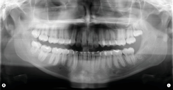
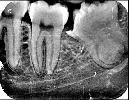
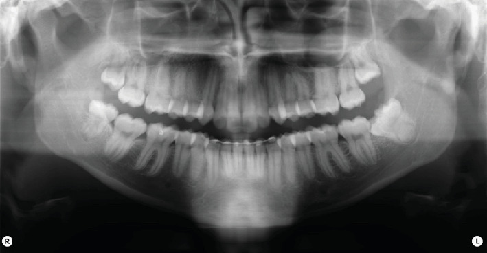
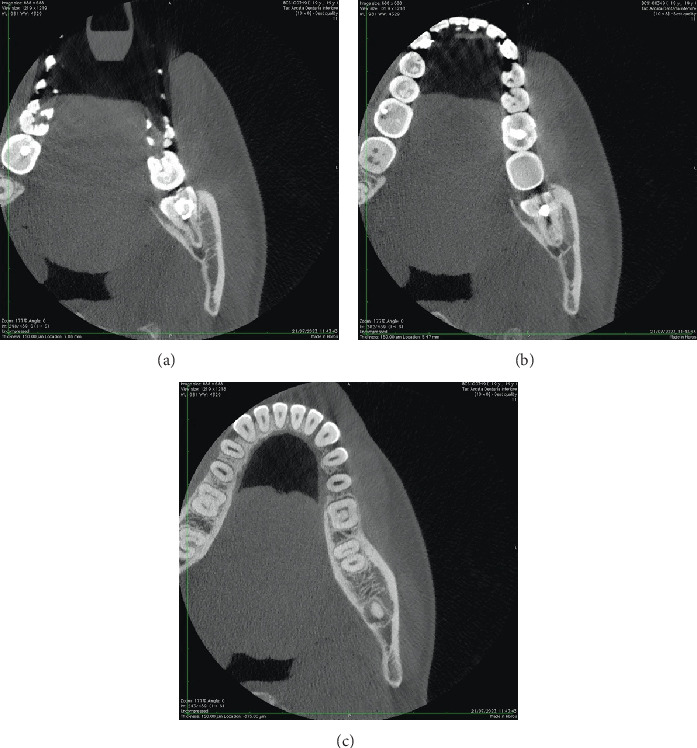
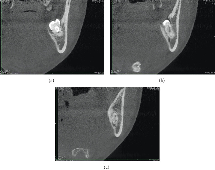
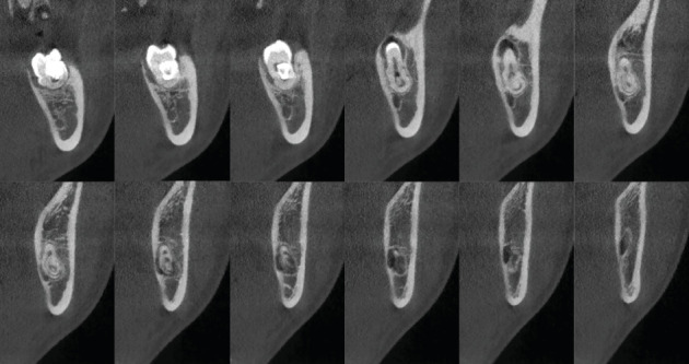
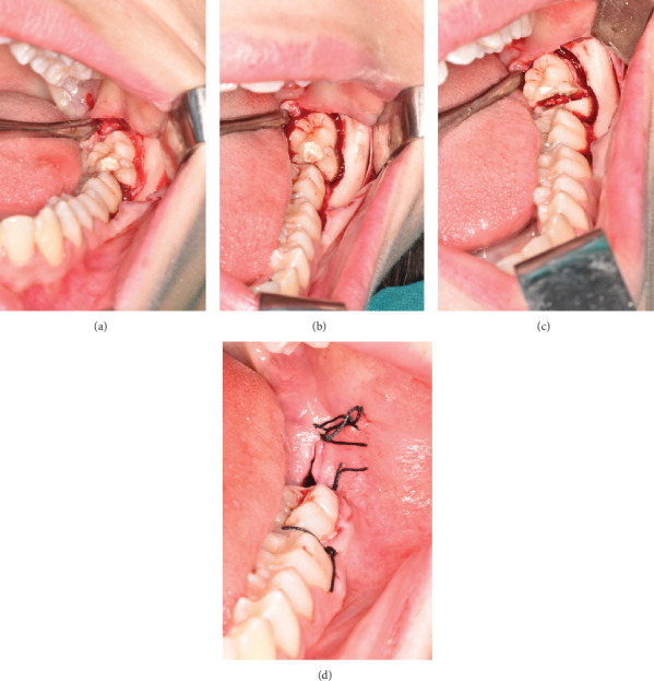
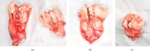
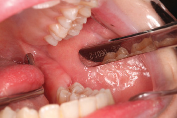
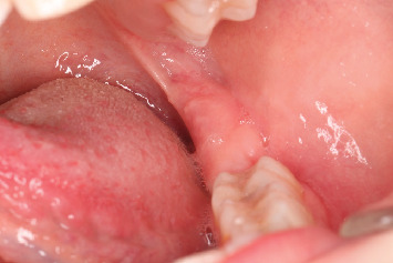
Gemination and fusion are rare developmental anomalies that can present significant diagnostic challenges. Due to the complexity of distinguishing between these conditions, the term "double tooth" is commonly employed in clinical practice. The precise etiology of these anomalies remains uncertain, and their occurrence in permanent dentition-particularly involving molars-is exceptionally rare. This report describes an uncommon case of gemination affecting the mandibular left third molar (tooth 3.8) and provides a comprehensive discussion contextualized within existing literature. The case report was prepared following the CARE guidelines to ensure methodological rigor and completeness. After an intraoral examination and radiographic assessment-including orthopantomography, periapical radiographs, and cone beam computed tomography (CBCT)-the patient underwent surgical extraction. The procedure involved administering a truncal nerve block to anesthetize the inferior alveolar and lingual nerves, supplemented by local infiltration anesthesia of the buccal nerve. A full-thickness mucoperiosteal flap was elevated, followed by ostectomy and odontotomy to facilitate extraction. The tooth was subsequently removed using a combination of elevators and forceps. Postoperative evaluations conducted at 1.5 and 3 months confirmed complete healing of the surgical site. A detailed analysis of pre- and postoperative radiographic and clinical findings validated the diagnosis of gemination, characterized by coronal continuity with a single root and root canal. Gemination of third molars is exceedingly rare, with only a few cases documented in the literature. To the best of our knowledge, this is the first reported instance of gemination involving the mandibular left third molar (tooth 3.8). This report contributes to the growing body of knowledge on developmental dental anomalies and highlights the importance of thorough differential diagnosis in similar clinical scenarios.
双生牙和融合牙是罕见的发育异常,会带来重大的诊断挑战。由于区分这些情况的复杂性,临床实践中通常使用“双牙”一词。这些异常的确切病因仍不确定,它们在恒牙列中出现,尤其是累及磨牙的情况极为罕见。本报告描述了一例罕见的下颌左第三磨牙(3.8 牙)双生牙病例,并结合现有文献进行了全面讨论。该病例报告是按照 CARE 指南编写的,以确保方法的严谨性和完整性。经过口腔内检查和影像学评估,包括全景片、根尖片和锥形束计算机断层扫描(CBCT)后,患者接受了手术拔除。手术过程包括对下牙槽神经和舌神经进行干神经阻滞麻醉,并辅以颊神经局部浸润麻醉。掀起全厚粘骨膜瓣,随后进行骨切除术和牙切开术以利于拔除。随后使用牙挺和牙钳拔除牙齿。术后 1.5 个月和 3 个月的评估证实手术部位完全愈合。对术前和术后影像学及临床检查结果的详细分析证实了双生牙的诊断,其特征为冠部连续且有单一牙根和根管。第三磨牙双生极为罕见,文献中仅有少数病例记载。据我们所知,这是首次报道的下颌左第三磨牙(3.8 牙)双生病例。本报告为有关发育性牙异常的知识体系增添了内容,并强调了在类似临床情况下进行全面鉴别诊断的重要性。