Dy Pauline Mae R, Yturralde Erick Martin H, Luna Jericho Thaddeus P
Department of Laboratories, Philippine General Hospital, University of the Philippines Manila.
Department of Obstetrics and Gynecology, Philippine General Hospital, University of the Philippines Manila.
Acta Med Philipp. 2025 Apr 30;59(5):82-87. doi: 10.47895/amp.vi0.10057. eCollection 2025.
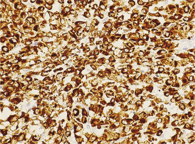
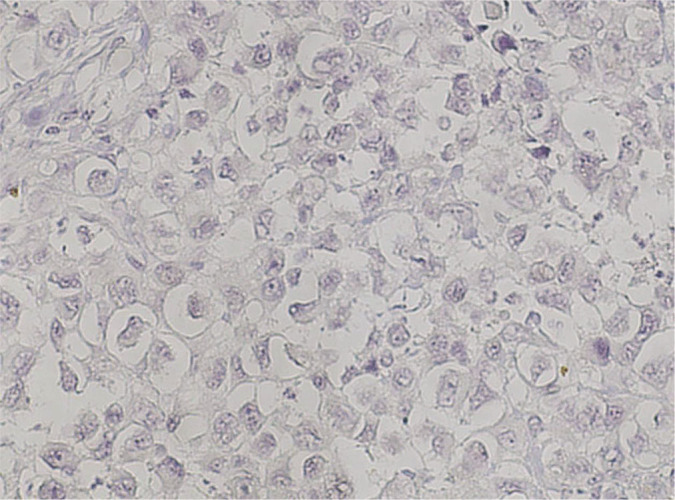
Epithelioid sarcoma is an uncommon mesenchymal malignancy which represents less than 1% of all sarcomas. Rarer still are reports of this tumor initially presenting in the vulva. We report a case of vulvar proximal-type epithelioid sarcoma. A 52-year-old had a 5-month history of slowly growing papule on the right labia majora. Excision of the mass revealed a tumor composed of large polygonal cells with abundant eosinophilic cytoplasm. An immunohistochemistry panel revealed cytokeratin AE1/AE3 positivity only. She underwent radical vulvectomy with bilateral groin node dissection. The specimen revealed a cream tan, firm, fairly defined mass at the right vulva. Microscopic examination showed a sheet-like growth pattern of large pleomorphic epithelioid cells with large vesicular nuclei and prominent nucleoli. The tumor showed loss of INI1 nuclear expression and absence of CD34 staining. EMA was positive. The case was signed out as proximal-type epithelioid sarcoma of the right vulva. Two months post-operatively, the patient was given concurrent chemotherapy with 5 cycles of cisplatin 40 mg/m and 6600 centigray vulvar intensity-modulated radiotherapy. She had no evidence of disease for five months until repeat workup showed tumor recurrence in the perineum. She was subsequently given 6 cycles of gemcitabine 900 mg/m and gemcitabine 900 mg/m with docetaxel 100 mg/m. Two months after, repeat workup showed persistent progressive disease in the vulva. She was subsequently given 4 cycles of doxorubicin 60 mg/m and is for repeat workup. The immunohistomorphologic features of this tumor, in addition to its unusual location, present a diagnostic challenge. Clues to the diagnosis include an initial presentation as a soft tissue mass and microscopic features showing the presence of epithelioid to spindle cytomorphology with an infiltrative growth pattern. Immunohistochemistry studies revealing the loss of INI1 nuclear expression and expression of epithelial markers would ultimately establish the diagnosis of this rare clinical entity.
上皮样肉瘤是一种罕见的间叶性恶性肿瘤,占所有肉瘤的比例不到1%。更罕见的是关于这种肿瘤最初在外阴出现的报道。我们报告一例外阴近端型上皮样肉瘤病例。一名52岁女性,右侧大阴唇有一个缓慢生长的丘疹,病史5个月。肿物切除显示肿瘤由大的多边形细胞组成,胞质丰富嗜酸性。免疫组化结果仅显示细胞角蛋白AE1/AE3阳性。她接受了根治性外阴切除术及双侧腹股沟淋巴结清扫术。标本显示右侧外阴有一个淡褐色、质地硬、边界较清晰的肿物。显微镜检查显示大片状生长的大的多形性上皮样细胞,核大呈泡状,核仁明显。肿瘤显示INI1核表达缺失及CD34染色阴性。EMA阳性。该病例诊断为右侧外阴近端型上皮样肉瘤。术后两个月,患者接受了顺铂40mg/m²共5个周期的同步化疗及6600厘戈瑞的外阴调强放疗。5个月内她没有疾病证据,直到复查显示会阴部肿瘤复发。随后她接受了吉西他滨900mg/m²共6个周期及吉西他滨900mg/m²联合多西他赛100mg/m²的治疗。两个月后,复查显示外阴持续进展性疾病。随后她接受了阿霉素60mg/m²共4个周期的治疗,以待复查。除了其不寻常的位置外,该肿瘤的免疫组织形态学特征也带来了诊断挑战。诊断线索包括最初表现为软组织肿物以及显微镜下特征显示存在上皮样至梭形细胞形态及浸润性生长模式。免疫组化研究显示INI1核表达缺失及上皮标志物表达最终将确立这种罕见临床实体的诊断。