• 文献检索
  • 文档翻译
  • 深度研究
  • 学术资讯
  • Suppr Zotero 插件Zotero 插件
  • 邀请有礼
  • 套餐&价格
  • 历史记录
应用&插件
Suppr Zotero 插件Zotero 插件浏览器插件Mac 客户端Windows 客户端微信小程序
定价
高级版会员购买积分包购买API积分包
服务
文献检索文档翻译深度研究API 文档MCP 服务
关于我们
关于 Suppr公司介绍联系我们用户协议隐私条款
关注我们

Suppr 超能文献

核心技术专利:CN118964589B侵权必究
粤ICP备2023148730 号-1Suppr @ 2026

文献检索

告别复杂PubMed语法,用中文像聊天一样搜索,搜遍4000万医学文献。AI智能推荐,让科研检索更轻松。

立即免费搜索

文件翻译

保留排版,准确专业,支持PDF/Word/PPT等文件格式,支持 12+语言互译。

免费翻译文档

深度研究

AI帮你快速写综述,25分钟生成高质量综述,智能提取关键信息,辅助科研写作。

立即免费体验

人内皮细胞通过Notch信号通路促进B型人神经干细胞表型。

Human endothelial cells promote a human neural stem cell type B phenotype via Notch signaling.

作者信息

Gutierrez Brenda, Liu Tzu Chia, Rodriguez Carly, Pastor-Alonso Oier, Lambing Hannah, Paredes Mercedes F, Flanagan Lisa A

机构信息

Department of Anatomy & Neurobiology, University of California Irvine, Irvine, CA, USA.

Sue & Bill Gross Stem Cell Research Center, University of California Irvine, Irvine, CA, USA.

出版信息

Nat Commun. 2025 May 30;16(1):5031. doi: 10.1038/s41467-025-60194-6.

DOI:10.1038/s41467-025-60194-6
PMID:40447618
原文链接:https://pmc.ncbi.nlm.nih.gov/articles/PMC12125299/
Abstract

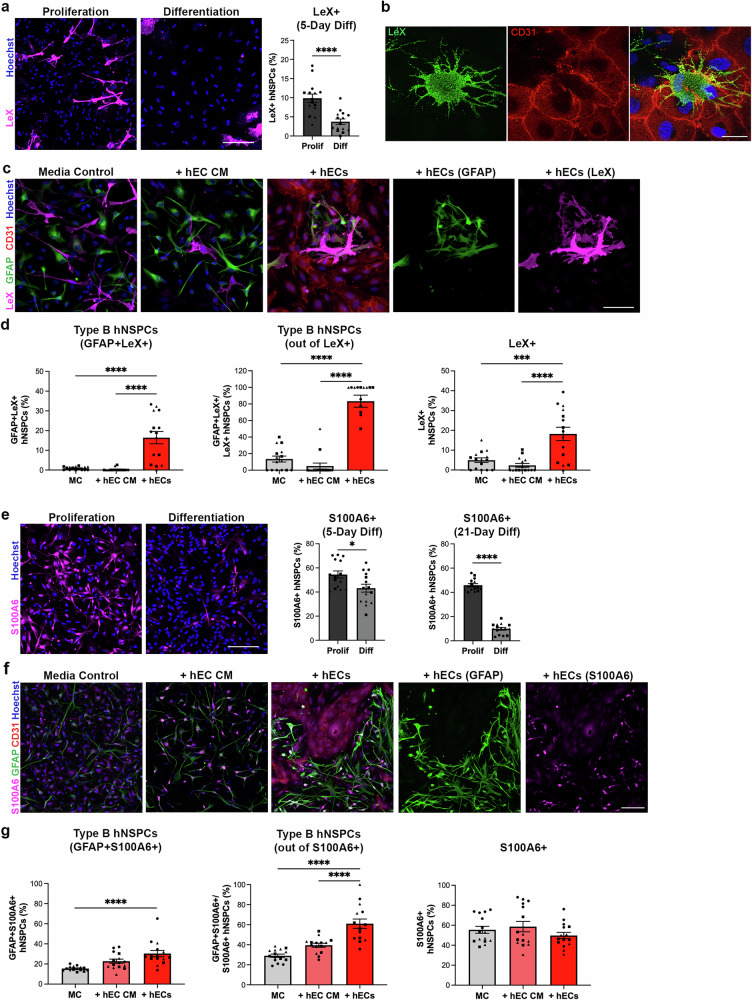
Neural stem and progenitor cell (NSPC) and vessel-forming endothelial cell (EC) communication throughout development and adulthood is vital for normal brain function. However, much remains unclear regarding coordinated regulation of these cells, particularly in humans. We find that contact with hECs increases hNSPC type B cells, which are GFAP-expressing adult NSPCs in the subventricular zone (SVZ), leading to generation of a human type B single-cell RNA sequencing (scRNAseq) dataset. Differential gene expression demonstrates an increase in Notch downstream mediators in type B hNSPCs after hEC contact. Blocking hNSPC Notch signaling, and reducing hEC expression of the Notch ligand DLL4, abrogates the effect of hECs on type B hNSPCs. We identify S100A6 and LeX as human type B cell markers, and analysis of the postnatal human SVZ confirms co-expression of GFAP, SOX2, S100A6, LeX and PROM1 in type B cells. Sites of contact are identified between type B hNSPCs and vasculature in the SVZ, providing evidence of human type B cell contact with hECs in the postnatal human brain. Thus, hEC contact promotes human type B cells via Notch signaling and these cells are in contact in stem cell niches in the human brain.

摘要

神经干细胞和祖细胞(NSPC)与血管形成内皮细胞(EC)在整个发育和成年期的通讯对于正常脑功能至关重要。然而,关于这些细胞的协调调控,尤其是在人类中,仍有许多不清楚的地方。我们发现与人类内皮细胞(hECs)接触会增加B型人类神经干细胞(hNSPCs),即脑室下区(SVZ)中表达胶质纤维酸性蛋白(GFAP)的成年神经干细胞,从而生成了一个人类B型单细胞RNA测序(scRNAseq)数据集。差异基因表达显示,hNSPCs与hECs接触后,B型hNSPCs中Notch下游介质增加。阻断hNSPC的Notch信号,并降低Notch配体DLL4的hEC表达,可消除hECs对B型hNSPCs的影响。我们将S100A6和LeX鉴定为人类B型细胞标志物,对出生后人脑SVZ的分析证实了GFAP、SOX2、S100A6、LeX和PROM1在B型细胞中的共表达。在SVZ中确定了B型hNSPCs与脉管系统之间的接触位点,为出生后人脑中B型细胞与hECs的接触提供了证据。因此,hEC接触通过Notch信号促进人类B型细胞,并且这些细胞在人类大脑的干细胞龛中存在接触。

https://cdn.ncbi.nlm.nih.gov/pmc/blobs/6c0f/12125299/edd2e13753e2/41467_2025_60194_Fig10_HTML.jpg
https://cdn.ncbi.nlm.nih.gov/pmc/blobs/6c0f/12125299/a5b52c41f138/41467_2025_60194_Fig1_HTML.jpg
https://cdn.ncbi.nlm.nih.gov/pmc/blobs/6c0f/12125299/a87867d25c55/41467_2025_60194_Fig2_HTML.jpg
https://cdn.ncbi.nlm.nih.gov/pmc/blobs/6c0f/12125299/08f0956640d8/41467_2025_60194_Fig3_HTML.jpg
https://cdn.ncbi.nlm.nih.gov/pmc/blobs/6c0f/12125299/d03941a3f6aa/41467_2025_60194_Fig4_HTML.jpg
https://cdn.ncbi.nlm.nih.gov/pmc/blobs/6c0f/12125299/a26a2aed5e5e/41467_2025_60194_Fig5_HTML.jpg
https://cdn.ncbi.nlm.nih.gov/pmc/blobs/6c0f/12125299/bb93ba6f2e14/41467_2025_60194_Fig6_HTML.jpg
https://cdn.ncbi.nlm.nih.gov/pmc/blobs/6c0f/12125299/66641b6f5602/41467_2025_60194_Fig7_HTML.jpg
https://cdn.ncbi.nlm.nih.gov/pmc/blobs/6c0f/12125299/8e880b1dbf1d/41467_2025_60194_Fig8_HTML.jpg
https://cdn.ncbi.nlm.nih.gov/pmc/blobs/6c0f/12125299/d6a275599aaf/41467_2025_60194_Fig9_HTML.jpg
https://cdn.ncbi.nlm.nih.gov/pmc/blobs/6c0f/12125299/edd2e13753e2/41467_2025_60194_Fig10_HTML.jpg

相似文献

1
Human endothelial cells promote a human neural stem cell type B phenotype via Notch signaling.人内皮细胞通过Notch信号通路促进B型人神经干细胞表型。
Nat Commun. 2025 May 30;16(1):5031. doi: 10.1038/s41467-025-60194-6.
2
TGF-β pro-oligodendrogenic effects on adult SVZ progenitor cultures and its interaction with the Notch signaling pathway.TGF-β 对成年 SVZ 祖细胞培养物的促少突胶质细胞生成作用及其与 Notch 信号通路的相互作用。
Glia. 2018 Feb;66(2):396-412. doi: 10.1002/glia.23253. Epub 2017 Oct 27.
3
Single-cell analysis of the ventricular-subventricular zone reveals signatures of dorsal and ventral adult neurogenesis.对脑室下区的单细胞分析揭示了背侧和腹侧成年神经发生的特征。
Elife. 2021 Jul 14;10:e67436. doi: 10.7554/eLife.67436.
4
Delta like 4 regulates cerebrovascular development and endothelial integrity via DLL4-NOTCH-CLDN5 pathway and is vulnerable to neonatal hyperoxia.Delta 样配体 4 通过 DLL4-NOTCH-CLDN5 通路调控脑血管发育和内皮完整性,且易受新生儿高氧的影响。
J Physiol. 2024 May;602(10):2265-2285. doi: 10.1113/JP285716. Epub 2024 Apr 17.
5
Evidence for a subventricular zone neural stem cell phagocytic activity stimulated by the vitamin K-dependent factor protein S.维生素K依赖因子蛋白S刺激脑室下区神经干细胞吞噬活性的证据。
Stem Cells. 2015 Feb;33(2):515-25. doi: 10.1002/stem.1862.
6
Feed-forward signaling by membrane-bound ligand receptor circuit: the case of NOTCH DELTA-like 4 ligand in endothelial cells.膜结合配体受体回路的正向信号转导:以内皮细胞中的 NOTCH DELTA 样 4 配体为例。
J Biol Chem. 2010 Dec 24;285(52):40681-9. doi: 10.1074/jbc.M110.176065. Epub 2010 Oct 19.
7
TRIP6 regulates neural stem cell maintenance in the postnatal mammalian subventricular zone.TRIP6调节出生后哺乳动物脑室下区神经干细胞的维持。
Dev Dyn. 2014 Sep;243(9):1130-42. doi: 10.1002/dvdy.24161. Epub 2014 Jul 21.
8
Fibrinogen induces neural stem cell differentiation into astrocytes in the subventricular zone via BMP signaling.纤维蛋白原通过 BMP 信号诱导侧脑室下区神经干细胞向星形胶质细胞分化。
Nat Commun. 2020 Jan 31;11(1):630. doi: 10.1038/s41467-020-14466-y.
9
Notch signaling in astrocytes and neuroblasts of the adult subventricular zone in health and after cortical injury.健康状态下及皮质损伤后成年脑室下区星形胶质细胞和成神经细胞中的Notch信号通路
Dev Neurosci. 2006;28(1-2):81-91. doi: 10.1159/000090755.
10
Ventricular-subventricular zone fractones are speckled basement membranes that function as a neural stem cell niche.室下区 fractones 是斑驳的基膜,作为神经干细胞龛起作用。
Mol Biol Cell. 2019 Jan 1;30(1):56-68. doi: 10.1091/mbc.E18-05-0286. Epub 2018 Oct 31.

本文引用的文献

1
Identity and nature of neural stem cells in the adult human subventricular zone.成体人类脑室下区神经干细胞的特性与本质
iScience. 2024 Feb 27;27(4):109342. doi: 10.1016/j.isci.2024.109342. eCollection 2024 Apr 19.
2
FOXG1 targets BMP repressors and cell cycle inhibitors in human neural progenitor cells.FOXG1 靶向人类神经祖细胞中的 BMP 抑制剂和细胞周期抑制剂。
Hum Mol Genet. 2023 Jul 20;32(15):2511-2522. doi: 10.1093/hmg/ddad089.
3
Cerebral neurovascular embryology, anatomic variations, and congenital brain arteriovenous lesions.
脑神经血管胚胎学、解剖变异与先天性脑动静脉病变。
J Neurointerv Surg. 2022 Sep;14(9):910-919. doi: 10.1136/neurintsurg-2021-018607. Epub 2022 Feb 15.
4
Cell cycle arrest determines adult neural stem cell ontogeny by an embryonic Notch-nonoscillatory Hey1 module.细胞周期阻滞通过胚胎 Notch-非振荡 Hey1 模块决定成年神经干细胞发生。
Nat Commun. 2021 Nov 12;12(1):6562. doi: 10.1038/s41467-021-26605-0.
5
An atlas of cortical arealization identifies dynamic molecular signatures.皮质区辨图集确定了动态分子特征。
Nature. 2021 Oct;598(7879):200-204. doi: 10.1038/s41586-021-03910-8. Epub 2021 Oct 6.
6
Single Cell Transcriptomics of Ependymal Cells Across Age, Region and Species Reveals Cilia-Related and Metal Ion Regulatory Roles as Major Conserved Ependymal Cell Functions.跨年龄、区域和物种的室管膜细胞单细胞转录组学揭示了纤毛相关和金属离子调节作用是室管膜细胞主要的保守功能。
Front Cell Neurosci. 2021 Jul 15;15:703951. doi: 10.3389/fncel.2021.703951. eCollection 2021.
7
Molecular architecture of the developing mouse brain.发育中老鼠大脑的分子结构。
Nature. 2021 Aug;596(7870):92-96. doi: 10.1038/s41586-021-03775-x. Epub 2021 Jul 28.
8
Single-cell analysis of the ventricular-subventricular zone reveals signatures of dorsal and ventral adult neurogenesis.对脑室下区的单细胞分析揭示了背侧和腹侧成年神经发生的特征。
Elife. 2021 Jul 14;10:e67436. doi: 10.7554/eLife.67436.
9
UCSC Cell Browser: visualize your single-cell data.UCSC Cell Browser:可视化您的单细胞数据。
Bioinformatics. 2021 Dec 7;37(23):4578-4580. doi: 10.1093/bioinformatics/btab503.
10
Integrated analysis of multimodal single-cell data.多模态单细胞数据的综合分析。
Cell. 2021 Jun 24;184(13):3573-3587.e29. doi: 10.1016/j.cell.2021.04.048. Epub 2021 May 31.