Yu Jiabin, Liu Qi, Xu Chenjie, Zhou Qinli, Xu Jiajun, Zhu Lingying, Chen Chen, Zhou Yahan, Xiao Binggang, Zheng Lin, Zhou Xiaofeng, Zhang Fengming, Ye Yuhang, Mi Hongmei, Zhang Dongping, Yang Li, Wu Zhiwei, Wang Jiayi, Chen Ming, Zhou Zhirui, Wang Haoyang, Wang Vicky Y, Wang Enyu, Xu Dong
Taizhou Campus, Zhejiang Cancer Hospital (Taizhou Cancer Hospital), Taizhou, Zhejiang, 317502, China.
College of Information Engineering, China Jiliang University, Hangzhou, Zhejiang, 310018, China.
BMC Med Inform Decis Mak. 2025 May 30;25(1):200. doi: 10.1186/s12911-025-03029-0.
This study aims to develop a deep learning framework that leverages modality imputation and subregion segmentation to improve grading accuracy in high-grade gliomas.
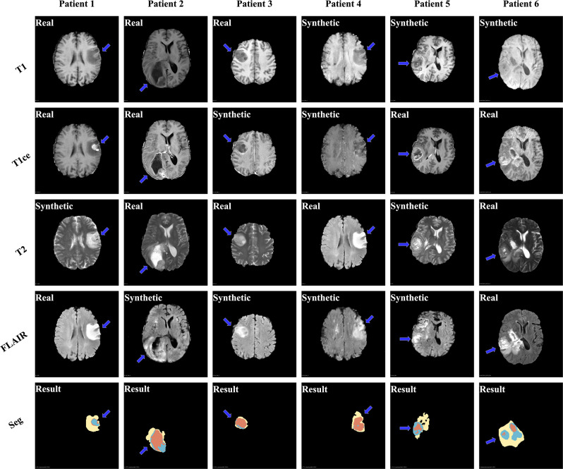
A retrospective analysis was conducted using data from 1,251 patients in the BraTS2021 dataset as the main cohort and 181 clinical cases collected from a medical center between April 2013 and June 2018 (51 years ± 17; 104 males) as the external test set. We propose a PatchGAN-based modality imputation network with an Aggregated Residual Transformer (ART) module combining Transformer self-attention and CNN feature extraction via residual links, paired with a U-Net variant for segmentation. Generative accuracy used PSNR and SSIM for modality conversions, while segmentation performance was measured with DSC and HD95 across necrotic core (NCR), edema (ED), and enhancing tumor (ET) regions. Senior radiologists conducted a comprehensive Likert-based assessment, with diagnostic accuracy evaluated by AUC. Statistical analysis was performed using the Wilcoxon signed-rank test and the DeLong test.
The best source-target modality pairs for imputation were T1 to T1ce and T1ce to T2 (p < 0.001). In subregion segmentation, the overall DSC was 0.878 and HD95 was 19.491, with the ET region showing the highest segmentation accuracy (DSC: 0.877, HD95: 12.149). Clinical validation revealed an improvement in grading accuracy by the senior radiologist, with the AUC increasing from 0.718 to 0.913 (P < 0.001) when using the combined imputation and segmentation models.
The proposed deep learning framework improves high-grade glioma grading by modality imputation and segmentation, aiding the senior radiologist and offering potential to advance clinical decision-making.
本研究旨在开发一种深度学习框架,该框架利用模态插补和子区域分割来提高高级别胶质瘤的分级准确性。
进行了一项回顾性分析,将来自BraTS2021数据集中1251例患者的数据作为主要队列,并将2013年4月至2018年6月期间从一个医疗中心收集的181例临床病例(51岁±17岁;104名男性)作为外部测试集。我们提出了一种基于PatchGAN的模态插补网络,该网络具有一个聚合残差Transformer(ART)模块,该模块通过残差链接将Transformer自注意力和CNN特征提取相结合,并与一个用于分割的U-Net变体配对。生成准确性使用PSNR和SSIM进行模态转换,而分割性能则通过坏死核心(NCR)、水肿(ED)和强化肿瘤(ET)区域的DSC和HD95进行测量。资深放射科医生进行了基于李克特量表的全面评估,诊断准确性通过AUC进行评估。使用Wilcoxon符号秩检验和DeLong检验进行统计分析。
用于插补的最佳源-目标模态对是T1到T1ce以及T1ce到T2(p < 0.001)。在子区域分割中,总体DSC为0.878,HD95为19.491,ET区域显示出最高的分割准确性(DSC:0.877,HD95:12.149)。临床验证表明资深放射科医生的分级准确性有所提高,使用联合插补和分割模型时,AUC从0.718增加到0.913(P < 0.001)。
所提出的深度学习框架通过模态插补和分割提高了高级别胶质瘤的分级,有助于资深放射科医生,并为推进临床决策提供了潜力。