• 文献检索
  • 文档翻译
  • 深度研究
  • 学术资讯
  • Suppr Zotero 插件Zotero 插件
  • 邀请有礼
  • 套餐&价格
  • 历史记录
应用&插件
Suppr Zotero 插件Zotero 插件浏览器插件Mac 客户端Windows 客户端微信小程序
定价
高级版会员购买积分包购买API积分包
服务
文献检索文档翻译深度研究API 文档MCP 服务
关于我们
关于 Suppr公司介绍联系我们用户协议隐私条款
关注我们

Suppr 超能文献

核心技术专利:CN118964589B侵权必究
粤ICP备2023148730 号-1Suppr @ 2026

文献检索

告别复杂PubMed语法,用中文像聊天一样搜索,搜遍4000万医学文献。AI智能推荐,让科研检索更轻松。

立即免费搜索

文件翻译

保留排版,准确专业,支持PDF/Word/PPT等文件格式,支持 12+语言互译。

免费翻译文档

深度研究

AI帮你快速写综述,25分钟生成高质量综述,智能提取关键信息,辅助科研写作。

立即免费体验

基于人工智能的胶质瘤分级以实现可靠诊断:提高可靠性的分析流程

AI-Based Glioma Grading for a Trustworthy Diagnosis: An Analytical Pipeline for Improved Reliability.

作者信息

Pitarch Carla, Ribas Vicent, Vellido Alfredo

机构信息

Computer Science Department, Universitat Politècnica de Catalunya (UPC), 08034 Barcelona, Spain.

Eurecat, Technology Centre of Catalonia, Digital Health Unit, 08005 Barcelona, Spain.

出版信息

Cancers (Basel). 2023 Jun 27;15(13):3369. doi: 10.3390/cancers15133369.

DOI:10.3390/cancers15133369
PMID:37444479
原文链接:https://pmc.ncbi.nlm.nih.gov/articles/PMC10341156/
Abstract

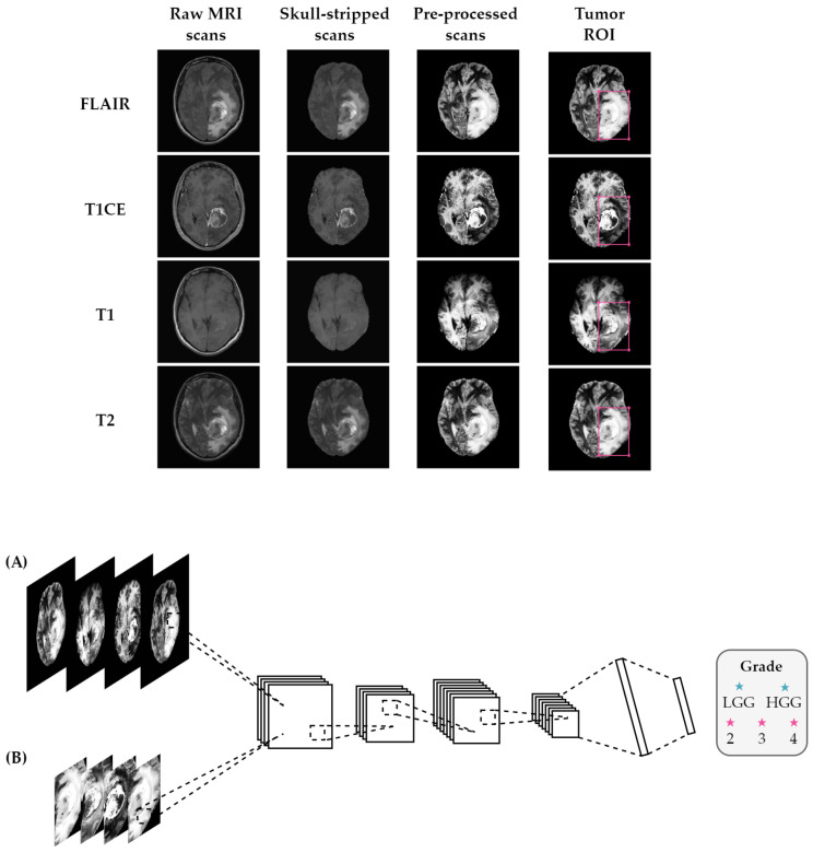
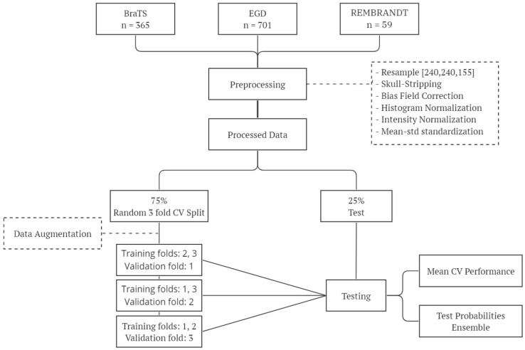
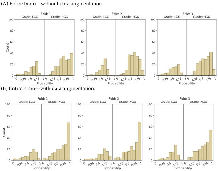
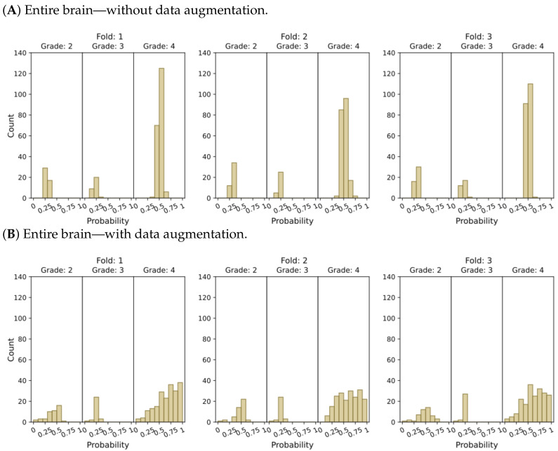
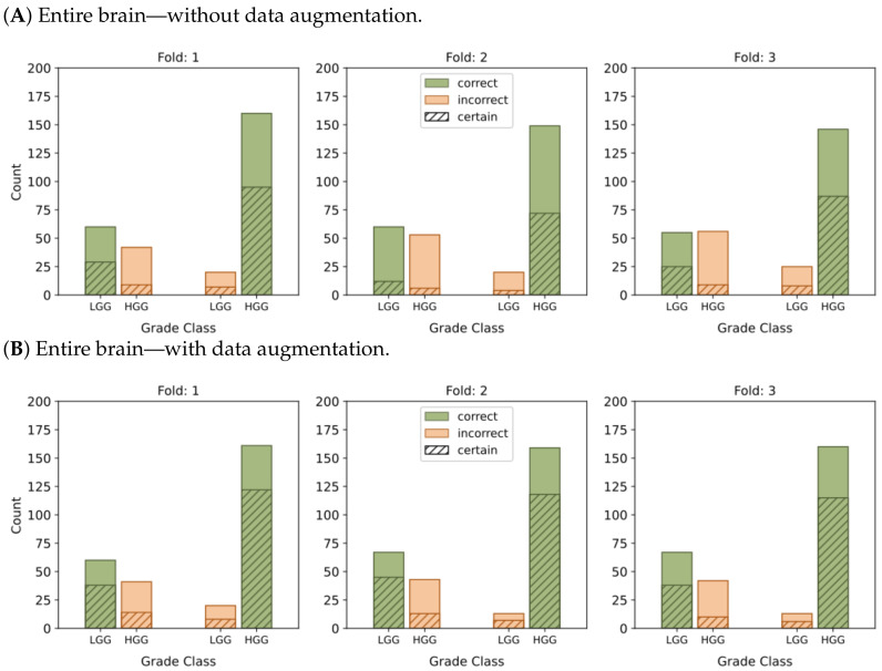
Glioma is the most common type of tumor in humans originating in the brain. According to the World Health Organization, gliomas can be graded on a four-stage scale, ranging from the most benign to the most malignant. The grading of these tumors from image information is a far from trivial task for radiologists and one in which they could be assisted by machine-learning-based decision support. However, the machine learning analytical pipeline is also fraught with perils stemming from different sources, such as inadvertent data leakage, adequacy of 2D image sampling, or classifier assessment biases. In this paper, we analyze a glioma database sourced from multiple datasets using a simple classifier, aiming to obtain a reliable tumor grading and, on the way, we provide a few guidelines to ensure such reliability. Our results reveal that by focusing on the tumor region of interest and using data augmentation techniques we significantly enhanced the accuracy and confidence in tumor classifications. Evaluation on an independent test set resulted in an AUC-ROC of 0.932 in the discrimination of low-grade gliomas from high-grade gliomas, and an AUC-ROC of 0.893 in the classification of grades 2, 3, and 4. The study also highlights the importance of providing, beyond generic classification performance, measures of how reliable and trustworthy the model's output is, thus assessing the model's certainty and robustness.

摘要

神经胶质瘤是人类起源于大脑的最常见肿瘤类型。根据世界卫生组织的分类,神经胶质瘤可分为四个等级,从最良性到最恶性。从图像信息中对这些肿瘤进行分级,对于放射科医生来说绝非易事,而基于机器学习的决策支持可以为他们提供帮助。然而,机器学习分析流程也充满了来自不同来源的风险,如无意的数据泄露、二维图像采样的充分性或分类器评估偏差。在本文中,我们使用一个简单的分类器分析了一个源自多个数据集的神经胶质瘤数据库,旨在获得可靠的肿瘤分级,在此过程中,我们提供了一些指导方针以确保这种可靠性。我们的结果表明,通过关注感兴趣的肿瘤区域并使用数据增强技术,我们显著提高了肿瘤分类的准确性和可信度。在独立测试集上的评估结果显示,在区分低级别神经胶质瘤和高级别神经胶质瘤时,曲线下面积(AUC-ROC)为0.932,在对2级、3级和4级进行分类时,AUC-ROC为0.893。该研究还强调了除了提供一般的分类性能外,还需提供模型输出的可靠性和可信度的衡量标准的重要性,从而评估模型的确定性和稳健性。

https://cdn.ncbi.nlm.nih.gov/pmc/blobs/fbbf/10341156/fd537534d44a/cancers-15-03369-g007a.jpg
https://cdn.ncbi.nlm.nih.gov/pmc/blobs/fbbf/10341156/983d82c7fa1f/cancers-15-03369-g001.jpg
https://cdn.ncbi.nlm.nih.gov/pmc/blobs/fbbf/10341156/188cd049d336/cancers-15-03369-g002.jpg
https://cdn.ncbi.nlm.nih.gov/pmc/blobs/fbbf/10341156/8af990ced39e/cancers-15-03369-g003a.jpg
https://cdn.ncbi.nlm.nih.gov/pmc/blobs/fbbf/10341156/ad562e4c10f6/cancers-15-03369-g004a.jpg
https://cdn.ncbi.nlm.nih.gov/pmc/blobs/fbbf/10341156/5c9132148b41/cancers-15-03369-g005a.jpg
https://cdn.ncbi.nlm.nih.gov/pmc/blobs/fbbf/10341156/b12763156e6e/cancers-15-03369-g006a.jpg
https://cdn.ncbi.nlm.nih.gov/pmc/blobs/fbbf/10341156/fd537534d44a/cancers-15-03369-g007a.jpg

相似文献

1
AI-Based Glioma Grading for a Trustworthy Diagnosis: An Analytical Pipeline for Improved Reliability.基于人工智能的胶质瘤分级以实现可靠诊断:提高可靠性的分析流程
Cancers (Basel). 2023 Jun 27;15(13):3369. doi: 10.3390/cancers15133369.
2
Voxel-based clustered imaging by multiparameter diffusion tensor images for glioma grading.基于体素的多参数弥散张量成像在胶质瘤分级中的聚类成像。
Neuroimage Clin. 2014 Aug 7;5:396-407. doi: 10.1016/j.nicl.2014.08.001. eCollection 2014.
3
Glioma grading using a machine-learning framework based on optimized features obtained from T perfusion MRI and volumes of tumor components.基于 T 灌注 MRI 优化特征和肿瘤成分体积的机器学习框架进行脑胶质瘤分级。
J Magn Reson Imaging. 2019 Oct;50(4):1295-1306. doi: 10.1002/jmri.26704. Epub 2019 Mar 20.
4
A quantitative model based on clinically relevant MRI features differentiates lower grade gliomas and glioblastoma.一种基于临床相关 MRI 特征的定量模型可区分低级别胶质瘤和胶质母细胞瘤。
Eur Radiol. 2020 Jun;30(6):3073-3082. doi: 10.1007/s00330-019-06632-8. Epub 2020 Feb 5.
5
Deriving quantitative information from multiparametric MRI via Radiomics: Evaluation of the robustness and predictive value of radiomic features in the discrimination of low-grade versus high-grade gliomas with machine learning.通过放射组学从多参数 MRI 中提取定量信息:利用机器学习评估放射组学特征在低级别与高级别胶质瘤鉴别中的稳健性和预测价值。
Phys Med. 2023 Mar;107:102538. doi: 10.1016/j.ejmp.2023.102538. Epub 2023 Feb 14.
6
Folic acid supplementation and malaria susceptibility and severity among people taking antifolate antimalarial drugs in endemic areas.在流行地区,服用抗叶酸抗疟药物的人群中,叶酸补充剂与疟疾易感性和严重程度的关系。
Cochrane Database Syst Rev. 2022 Feb 1;2(2022):CD014217. doi: 10.1002/14651858.CD014217.
7
A comparative study for glioma classification using deep convolutional neural networks.使用深度卷积神经网络进行胶质瘤分类的比较研究。
Math Biosci Eng. 2021 Jan 29;18(2):1550-1572. doi: 10.3934/mbe.2021080.
8
Automated discrimination of lower and higher grade gliomas based on histopathological image analysis.基于组织病理学图像分析的低级别和高级别胶质瘤自动鉴别
J Pathol Inform. 2015 Mar 24;6:15. doi: 10.4103/2153-3539.153914. eCollection 2015.
9
Optimizing a machine learning based glioma grading system using multi-parametric MRI histogram and texture features.利用多参数MRI直方图和纹理特征优化基于机器学习的胶质瘤分级系统。
Oncotarget. 2017 Jul 18;8(29):47816-47830. doi: 10.18632/oncotarget.18001.
10
Measurements of diagnostic examination performance using quantitative apparent diffusion coefficient and proton MR spectroscopic imaging in the preoperative evaluation of tumor grade in cerebral gliomas.使用定量表观扩散系数和质子磁共振波谱成像对脑胶质瘤术前肿瘤分级进行诊断检查性能的测量。
Eur J Radiol. 2011 Nov;80(2):462-70. doi: 10.1016/j.ejrad.2010.07.017. Epub 2010 Aug 13.

引用本文的文献

1
Advanced MRI, Radiomics and Radiogenomics in Unravelling Incidental Glioma Grading and Genetic Status: Where Are We?高级磁共振成像、影像组学和放射基因组学在解读偶然发现的胶质瘤分级和基因状态中的应用:我们目前的进展如何?
Medicina (Kaunas). 2025 Aug 12;61(8):1453. doi: 10.3390/medicina61081453.
2
Hybrid classical and quantum computing for enhanced glioma tumor classification using TCGA data.利用TCGA数据的混合经典与量子计算用于增强胶质瘤肿瘤分类
Sci Rep. 2025 Jul 17;15(1):25935. doi: 10.1038/s41598-025-97067-3.
3
Artificial intelligence in cancer pathology: Applications, challenges, and future directions.

本文引用的文献

1
Combined molecular subtyping, grading, and segmentation of glioma using multi-task deep learning.使用多任务深度学习对胶质瘤进行联合分子亚型、分级和分割。
Neuro Oncol. 2023 Feb 14;25(2):279-289. doi: 10.1093/neuonc/noac166.
2
Enhancing the REMBRANDT MRI collection with expert segmentation labels and quantitative radiomic features.利用专家分割标签和定量放射组学特征增强 REMBRANDT MRI 数据集。
Sci Data. 2022 Jun 14;9(1):338. doi: 10.1038/s41597-022-01415-1.
3
Advancements in Oncology with Artificial Intelligence-A Review Article.人工智能在肿瘤学中的进展——一篇综述文章
癌症病理学中的人工智能:应用、挑战及未来方向。
Cytojournal. 2025 Apr 19;22:45. doi: 10.25259/Cytojournal_272_2024. eCollection 2025.
4
Deep learning-driven modality imputation and subregion segmentation to enhance high-grade glioma grading.深度学习驱动的模态插补和子区域分割以提高高级别胶质瘤分级。
BMC Med Inform Decis Mak. 2025 May 30;25(1):200. doi: 10.1186/s12911-025-03029-0.
5
Evolving and Novel Applications of Artificial Intelligence in Cancer Imaging.人工智能在癌症成像中的不断发展与新应用
Cancers (Basel). 2025 Apr 30;17(9):1510. doi: 10.3390/cancers17091510.
6
Revolutionizing Radiology With Artificial Intelligence.用人工智能革新放射学。
Cureus. 2024 Oct 29;16(10):e72646. doi: 10.7759/cureus.72646. eCollection 2024 Oct.
7
End-to-End Multi-task Learning Architecture for Brain Tumor Analysis with Uncertainty Estimation in MRI Images.端到端多任务学习架构,用于 MRI 图像中的脑肿瘤分析及不确定性估计。
J Imaging Inform Med. 2024 Oct;37(5):2149-2172. doi: 10.1007/s10278-024-01009-w. Epub 2024 Apr 2.
8
Advances in the Use of Deep Learning for the Analysis of Magnetic Resonance Image in Neuro-Oncology.深度学习在神经肿瘤学磁共振图像分析中的应用进展。
Cancers (Basel). 2024 Jan 10;16(2):300. doi: 10.3390/cancers16020300.
Cancers (Basel). 2022 Mar 6;14(5):1349. doi: 10.3390/cancers14051349.
4
Developing and validating a deep learning and radiomic model for glioma grading using multiplanar reconstructed magnetic resonance contrast-enhanced T1-weighted imaging: a robust, multi-institutional study.使用多平面重建磁共振对比增强T1加权成像开发并验证用于胶质瘤分级的深度学习和放射组学模型:一项稳健的多机构研究
Quant Imaging Med Surg. 2022 Feb;12(2):1517-1528. doi: 10.21037/qims-21-722.
5
Deep Learning for Brain Tumor Segmentation: A Survey of State-of-the-Art.用于脑肿瘤分割的深度学习:最新技术综述
J Imaging. 2021 Jan 29;7(2):19. doi: 10.3390/jimaging7020019.
6
The 2021 WHO Classification of Tumors of the Central Nervous System: a summary.2021 年世卫组织中枢神经系统肿瘤分类:概述。
Neuro Oncol. 2021 Aug 2;23(8):1231-1251. doi: 10.1093/neuonc/noab106.
7
The Erasmus Glioma Database (EGD): Structural MRI scans, WHO 2016 subtypes, and segmentations of 774 patients with glioma.伊拉斯谟胶质瘤数据库(EGD):774例胶质瘤患者的结构磁共振成像扫描、2016年世界卫生组织亚型及分割图像
Data Brief. 2021 Jun 2;37:107191. doi: 10.1016/j.dib.2021.107191. eCollection 2021 Aug.
8
Multiclass magnetic resonance imaging brain tumor classification using artificial intelligence paradigm.使用人工智能范式的多类别磁共振成像脑肿瘤分类
Comput Biol Med. 2020 Jul;122:103804. doi: 10.1016/j.compbiomed.2020.103804. Epub 2020 May 30.
9
Brain extraction on MRI scans in presence of diffuse glioma: Multi-institutional performance evaluation of deep learning methods and robust modality-agnostic training.MRI 扫描中存在弥漫性胶质瘤时的脑提取:深度学习方法的多机构性能评估和稳健的模态不可知训练。
Neuroimage. 2020 Oct 15;220:117081. doi: 10.1016/j.neuroimage.2020.117081. Epub 2020 Jun 27.
10
Automated glioma grading on conventional MRI images using deep convolutional neural networks.使用深度卷积神经网络对传统MRI图像进行自动脑胶质瘤分级
Med Phys. 2020 Jul;47(7):3044-3053. doi: 10.1002/mp.14168. Epub 2020 May 11.