Yan Zhong-Jiang, Ye Maosen, Li Jiexi, Zhang Deng-Feng, Yao Yong-Gang
State Key Laboratory of Genetic Evolution & Animal Models, Yunnan Key Laboratory of Animal Models and Human Disease Mechanisms, and KIZ-CUHK Joint Laboratory of Bioresources and Molecular Research in Common Diseases, Kunming Institute of Zoology, Chinese Academy of Sciences, Kunming, Yunnan, 650204, China.
Kunming College of Life Science, University of Chinese Academy of Sciences, Kunming, Yunnan, 650204, China.
Mol Neurodegener. 2025 May 31;20(1):62. doi: 10.1186/s13024-025-00853-w.
Alzheimer's disease (AD) is a progressive neurodegenerative disorder characterized by the accumulation of amyloid-β plaques, tau hyperphosphorylation, and neuroinflammation. The choroid plexus (ChP), serving as the blood-cerebrospinal fluid-brain barrier, plays essential roles in immune response to stress and brain homeostasis. However, the cellular and molecular contributions of the ChP to AD progression remain inadequately understood.
To elucidate the molecular abnormalities during the early stages of AD, we acquired single-cell transcription profiling of ChP from APP/PS1 mice with early-stage of Aβ pathology and litter-mate controls. The transcriptional alterations that occurred in each cell type were identified by differentially expressed genes, cell-cell communications and pseudotemporal trajectory analysis. The findings were subsequently validated by a series of in situ and in vitro assays.
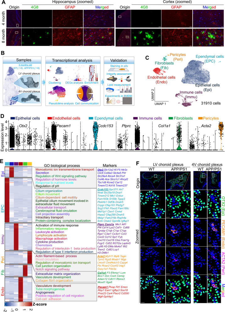
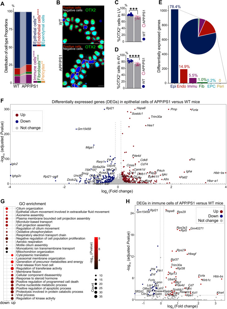
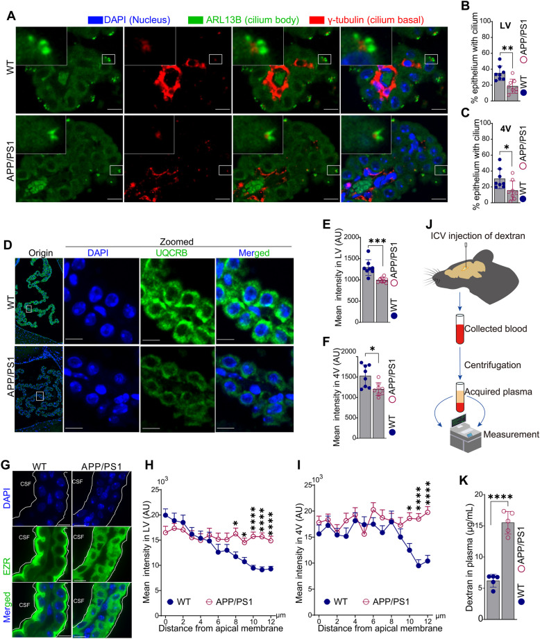
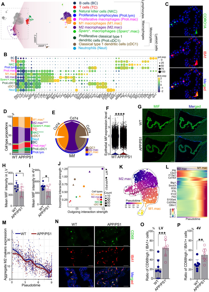
We constructed a comprehensive atlas of ChP at single-cell resolution and identified six major cell types and immune subclusters in male mice. The majority of dysregulated genes were found in the epithelial cells of APP/PS1 mice in comparison to wild-type (WT) mice, and most of these genes belonged to down-regulated module involved in mitochondrial respirasome assembly, cilium organization, and barrier integrity. The disruption of the epithelial barrier resulted in the downregulation of macrophage migration inhibitory factor (MIF) secretion in APP/PS1 mice, leading to macrophage activation and increased phagocytosis of Aβ. Concurrently, ligands (e.g., APOE) secreted by macrophages and other ChP cells facilitated the entry of lipids into ependymal cells, leading to lipid accumulation and the activation of microglia in the brain parenchyma in APP/PS1 mice compared to WT controls.
Taken together, these data profiled early transcriptional and cellular abnormalities of ChP within an AD mouse model, providing novel insights of cerebral vasculature into the pathobiology of AD.
阿尔茨海默病(AD)是一种进行性神经退行性疾病,其特征为β淀粉样蛋白斑块的积累、tau蛋白过度磷酸化和神经炎症。脉络丛(ChP)作为血脑脊液屏障,在对应激的免疫反应和脑内环境稳定中发挥着重要作用。然而,脉络丛对AD进展的细胞和分子贡献仍未得到充分了解。
为了阐明AD早期阶段的分子异常,我们获取了具有早期Aβ病理特征的APP/PS1小鼠及其同窝对照小鼠脉络丛的单细胞转录谱。通过差异表达基因、细胞间通讯和拟时间轨迹分析确定了每种细胞类型中发生的转录变化。随后通过一系列原位和体外试验对这些发现进行了验证。
我们构建了雄性小鼠脉络丛的单细胞分辨率综合图谱,并确定了六种主要细胞类型和免疫亚群。与野生型(WT)小鼠相比,APP/PS1小鼠的上皮细胞中发现了大多数失调基因,其中大多数基因属于下调模块,参与线粒体呼吸体组装、纤毛组织和屏障完整性。上皮屏障的破坏导致APP/PS1小鼠中巨噬细胞迁移抑制因子(MIF)分泌下调,从而导致巨噬细胞活化和Aβ吞噬增加。同时,与WT对照相比,巨噬细胞和其他脉络丛细胞分泌的配体(如APOE)促进脂质进入室管膜细胞,导致脂质积累和APP/PS1小鼠脑实质中小胶质细胞的活化。
综上所述,这些数据描绘了AD小鼠模型中脉络丛早期转录和细胞异常情况,为脑血管系统对AD病理生物学的研究提供了新的见解。