Nistor Irina Manuela, Fica Simona, Martin Sorina Carmen, Mitrache Marius Lucian, Oprea Theodor Eugen, Sirbu Anca Elena, Barbu Carmen Gabriela
Department of Endocrinology, University of Medicine and Pharmacy "Carol Davila," Bucharest, Romania.
Department of Endocrinology, University of Medicine and Pharmacy "Carol Davila," Dionisie Lupu Street No. 37, Bucharest 030167, Romania.
Ther Adv Endocrinol Metab. 2025 May 29;16:20420188251332082. doi: 10.1177/20420188251332082. eCollection 2025.
Although measuring bone mineral density (BMD) with dual X-ray absorptiometry (DXA) represents the standard of diagnosis and management of osteoporosis, there is a significant number of fragility fractures occurring in young patients without low BMD. Recently, clinical risk tools included hip axis length (HAL), a geometric parameter derived from the hip DXA scan, as a predictor of hip fractures in older postmenopausal women. This study aims to evaluate the relationship between HAL and other cortical bone fractures in young postmenopausal, clinically healthy women.
This study is a retrospective analysis of Lunar DXA scans of 206 normal or overweight Caucasian women aged 40-60, who had less than 10 years of menopause without secondary causes of osteoporosis, no prior osteoporosis diagnosis or medication, and no history of hip or vertebral fractures.
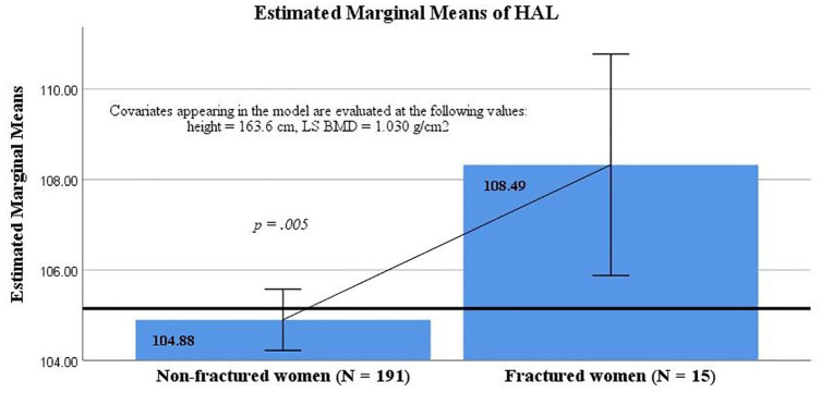
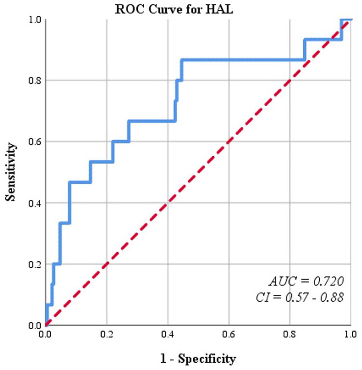
The 15 fractured women displayed statistically greater HAL values compared to the 191 non-fractured subjects (109.43 ± 6.44 vs 104.81 ± 5.32 mm, = 0.002), even though there were no significant differences in age, body mass index, or BMD. The difference in HAL remained significant after adjusting for lumbar spine (LS) BMD and height (108.49 ± 1.23 vs 104.88 ± 0.34 mm, = 0.005). HAL proved to be a fair indicator of non-hip, non-vertebral cortical fractures (area under curve = 0.720, = 0.003), with a sensitivity of 86.7% and a specificity of 55.5%.
HAL was positively associated with non-hip, non-vertebral cortical bone fragility fractures in young postmenopausal, clinically healthy women and had significantly greater values in the fractured subgroup even after adjusting for LS BMD and height.
尽管采用双能X线吸收法(DXA)测量骨密度(BMD)是骨质疏松症诊断和管理的标准方法,但仍有相当数量的年轻患者在骨密度未降低的情况下发生脆性骨折。最近,临床风险评估工具将髋轴长度(HAL)纳入其中,HAL是一种从髋部DXA扫描得出的几何参数,可作为老年绝经后女性髋部骨折的预测指标。本研究旨在评估HAL与年轻绝经后临床健康女性其他皮质骨骨折之间的关系。
本研究对206名年龄在40 - 60岁的正常或超重白种女性的Lunar DXA扫描结果进行回顾性分析。这些女性绝经时间少于10年,无骨质疏松症继发原因,既往未诊断出骨质疏松症或接受过相关治疗且无髋部或椎体骨折史。
与191名未骨折的受试者相比,15名骨折女性的HAL值在统计学上显著更高(分别为109.43 ± 6.44与104.81 ± 5.32毫米;P = 0.002),尽管在年龄、体重指数或骨密度方面无显著差异。在对腰椎(LS)骨密度和身高进行校正后,HAL的差异仍然显著(分别为108.49 ± 1.23与104.88 ± 0.34毫米;P = 0.005)。HAL被证明是髋部和椎体以外皮质骨骨折(曲线下面积 = 0.720;P = 0.003)的一个有效指标,敏感性为86.7%,特异性为55.5%。
在年轻绝经后临床健康女性中HAL与髋部和椎体以外皮质骨脆性骨折呈正相关,即使在对LS骨密度和身高进行校正后,骨折亚组中的HAL值仍显著更高。