• 文献检索
  • 文档翻译
  • 深度研究
  • 学术资讯
  • Suppr Zotero 插件Zotero 插件
  • 邀请有礼
  • 套餐&价格
  • 历史记录
应用&插件
Suppr Zotero 插件Zotero 插件浏览器插件Mac 客户端Windows 客户端微信小程序
定价
高级版会员购买积分包购买API积分包
服务
文献检索文档翻译深度研究API 文档MCP 服务
关于我们
关于 Suppr公司介绍联系我们用户协议隐私条款
关注我们

Suppr 超能文献

核心技术专利:CN118964589B侵权必究
粤ICP备2023148730 号-1Suppr @ 2026

文献检索

告别复杂PubMed语法,用中文像聊天一样搜索,搜遍4000万医学文献。AI智能推荐,让科研检索更轻松。

立即免费搜索

文件翻译

保留排版,准确专业,支持PDF/Word/PPT等文件格式,支持 12+语言互译。

免费翻译文档

深度研究

AI帮你快速写综述,25分钟生成高质量综述,智能提取关键信息,辅助科研写作。

立即免费体验

在斑马鱼生长突增期间,胆管细胞在部分肝损伤后对肝细胞再生有贡献。

Cholangiocytes contribute to hepatocyte regeneration after partial liver injury during growth spurt in zebrafish.

作者信息

Eski Sema Elif, Mi Jiarui, Pozo-Morales Macarena, Hovhannisyan Gabriel Garnik, Perazzolo Camille, Manco Rita, Ez-Zammoury Imane, Barbhaya Dev, Lefort Anne, Libert Frédérick, Marini Federico, Gurzov Esteban N, Andersson Olov, Singh Sumeet Pal

机构信息

Laboratory of Regeneration and Stress Biology, Institut de Recherche Interdisciplinaire en Biologie Humaine et Moléculaire (IRIBHM-Jacques E. Dumont), Université libre de Bruxelles, Brussels, Belgium.

Department of Cell and Molecular Biology, Karolinska Institutet, 17177, Stockholm, Sweden.

出版信息

Nat Commun. 2025 Jun 6;16(1):5260. doi: 10.1038/s41467-025-60334-y.

DOI:10.1038/s41467-025-60334-y
PMID:40480975
原文链接:https://pmc.ncbi.nlm.nih.gov/articles/PMC12144294/
Abstract

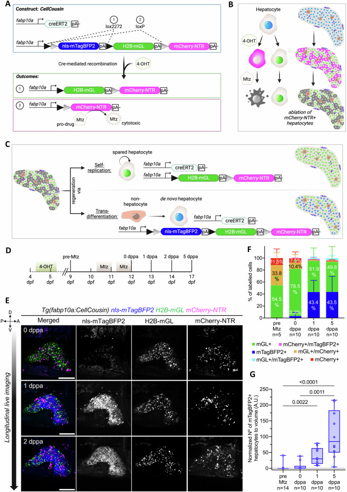
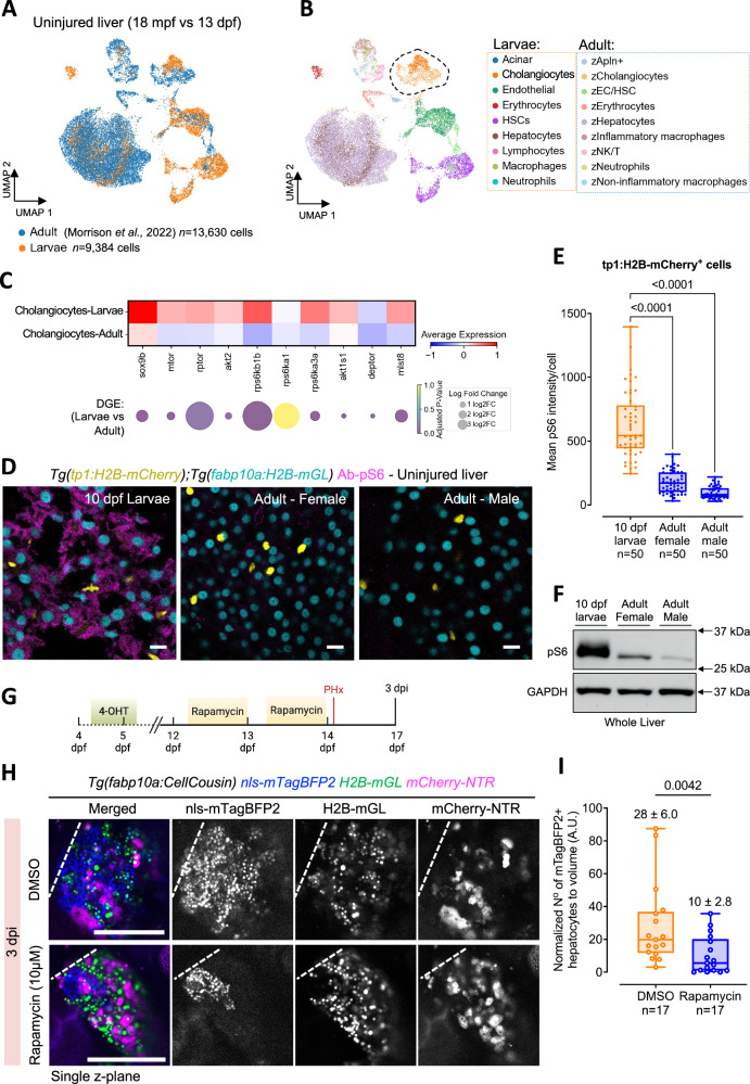
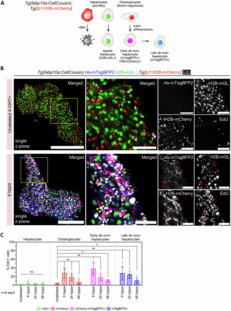
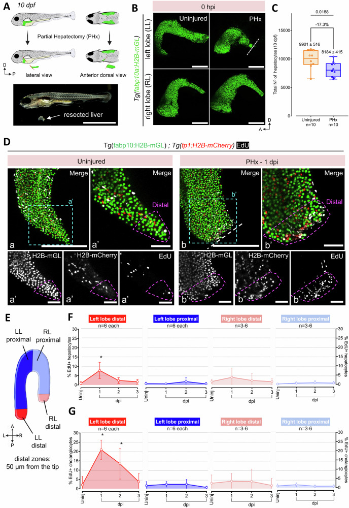
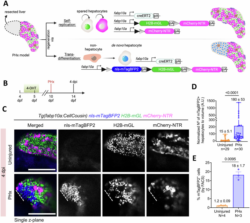
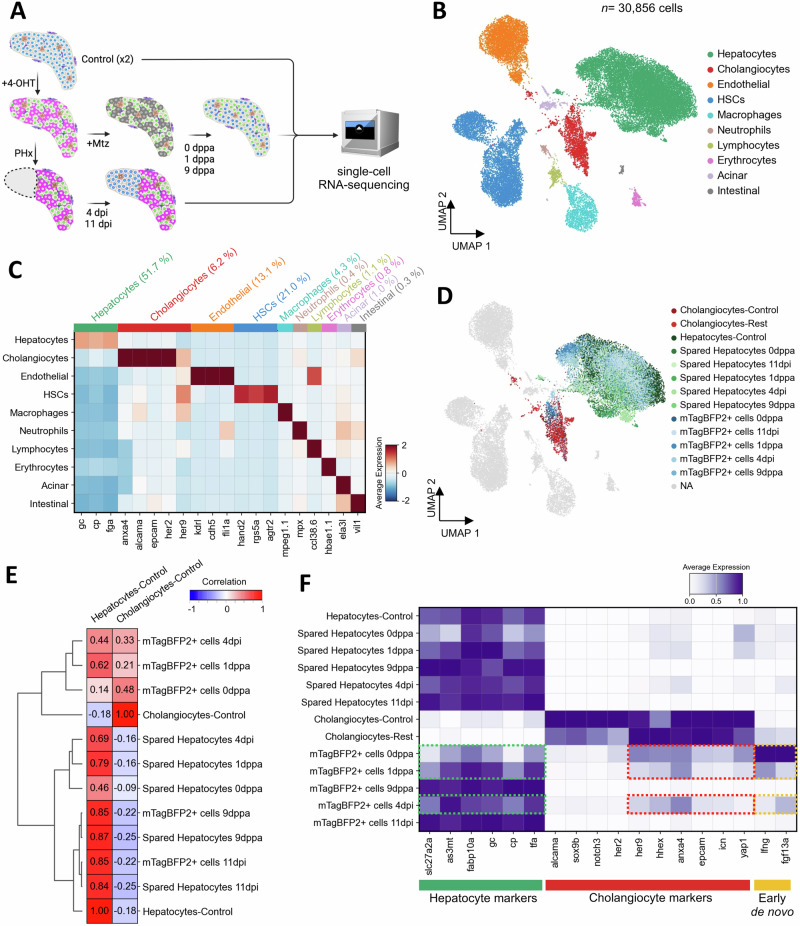
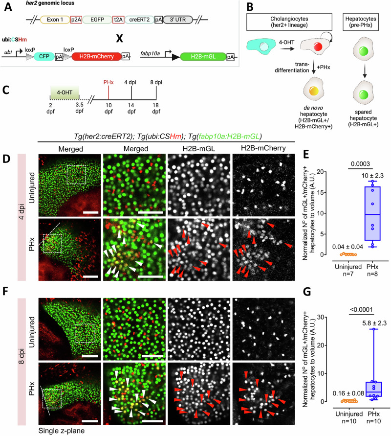
The liver's regenerative ability depends on injury extent. Minor injuries are repaired by hepatocyte self-duplication, while severe damage triggers cholangiocyte involvement in hepatocyte recovery. This paradigm is well-documented for adult animals but is less explored during rapid growth. We design two partial liver injury models in zebrafish, which were investigated during growth spurts: 1) partial ablation, killing half the hepatocytes; and 2) partial hepatectomy, removing half a liver lobe. In both injuries, de novo hepatocytes emerged alongside existing ones. Single-cell transcriptomics and lineage tracing with Cre-driver lines generated by genome editing identified cholangiocytes as the source of de novo hepatocytes. We further identify active mTORC1 signalling in the uninjured liver of growing animal to be a regulator of the enhanced plasticity of cholangiocytes. Our study suggests cholangiocyte-to-hepatocyte transdifferentiation as the primary mechanism of liver regeneration during periods of rapid growth.

摘要

肝脏的再生能力取决于损伤程度。轻微损伤通过肝细胞自我复制修复,而严重损伤则会促使胆管细胞参与肝细胞的恢复。这种模式在成年动物中已有充分记录,但在快速生长过程中研究较少。我们在斑马鱼中设计了两种部分肝损伤模型,并在生长高峰期对其进行研究:1)部分消融,杀死一半的肝细胞;2)部分肝切除术,切除半个肝叶。在这两种损伤中,新生肝细胞与现有的肝细胞一同出现。单细胞转录组学以及通过基因组编辑产生的Cre驱动系进行的谱系追踪将胆管细胞确定为新生肝细胞的来源。我们进一步确定,生长中动物未受伤肝脏中活跃的mTORC1信号是胆管细胞增强可塑性的调节因子。我们的研究表明,胆管细胞向肝细胞的转分化是快速生长期间肝脏再生的主要机制。

https://cdn.ncbi.nlm.nih.gov/pmc/blobs/f43b/12144294/e3dbdac80b32/41467_2025_60334_Fig7_HTML.jpg
https://cdn.ncbi.nlm.nih.gov/pmc/blobs/f43b/12144294/c78e55b71f04/41467_2025_60334_Fig1_HTML.jpg
https://cdn.ncbi.nlm.nih.gov/pmc/blobs/f43b/12144294/685a1865fe4a/41467_2025_60334_Fig2_HTML.jpg
https://cdn.ncbi.nlm.nih.gov/pmc/blobs/f43b/12144294/53e35ebab0fe/41467_2025_60334_Fig3_HTML.jpg
https://cdn.ncbi.nlm.nih.gov/pmc/blobs/f43b/12144294/e32223929bd7/41467_2025_60334_Fig4_HTML.jpg
https://cdn.ncbi.nlm.nih.gov/pmc/blobs/f43b/12144294/bc401c241a06/41467_2025_60334_Fig5_HTML.jpg
https://cdn.ncbi.nlm.nih.gov/pmc/blobs/f43b/12144294/0e7d22afc095/41467_2025_60334_Fig6_HTML.jpg
https://cdn.ncbi.nlm.nih.gov/pmc/blobs/f43b/12144294/e3dbdac80b32/41467_2025_60334_Fig7_HTML.jpg

相似文献

1
Cholangiocytes contribute to hepatocyte regeneration after partial liver injury during growth spurt in zebrafish.在斑马鱼生长突增期间,胆管细胞在部分肝损伤后对肝细胞再生有贡献。
Nat Commun. 2025 Jun 6;16(1):5260. doi: 10.1038/s41467-025-60334-y.
2
Regeneration of liver after extreme hepatocyte loss occurs mainly via biliary transdifferentiation in zebrafish.在斑马鱼中,极端肝细胞损失后的肝脏再生主要通过胆管细胞转分化发生。
Gastroenterology. 2014 Mar;146(3):789-800.e8. doi: 10.1053/j.gastro.2013.11.045. Epub 2013 Dec 4.
3
Mammalian Target of Rapamycin Complex 1 Signaling Is Required for the Dedifferentiation From Biliary Cell to Bipotential Progenitor Cell in Zebrafish Liver Regeneration.哺乳动物雷帕霉素靶蛋白复合物 1 信号通路在斑马鱼肝脏再生中胆管细胞向双潜能祖细胞去分化过程中发挥作用。
Hepatology. 2019 Dec;70(6):2092-2106. doi: 10.1002/hep.30790. Epub 2019 Aug 12.
4
Progranulin A Promotes Compensatory Hepatocyte Proliferation via HGF/c-Met Signaling after Partial Hepatectomy in Zebrafish.颗粒蛋白前体 A 通过 HGF/c-Met 信号促进斑马鱼部分肝切除后的代偿性肝细胞增殖。
Int J Mol Sci. 2021 Oct 18;22(20):11217. doi: 10.3390/ijms222011217.
5
De novo formation of the biliary system by TGFβ-mediated hepatocyte transdifferentiation.TGFβ 介导体肝细胞转分化形成新的胆道系统。
Nature. 2018 May;557(7704):247-251. doi: 10.1038/s41586-018-0075-5. Epub 2018 May 2.
6
Extensive conversion of hepatic biliary epithelial cells to hepatocytes after near total loss of hepatocytes in zebrafish.在斑马鱼中,近全量肝细胞丢失后,肝脏胆管上皮细胞广泛转化为肝细胞。
Gastroenterology. 2014 Mar;146(3):776-88. doi: 10.1053/j.gastro.2013.10.019. Epub 2013 Oct 19.
7
Hepatocyte-to-cholangiocyte conversion occurs through transdifferentiation independently of proliferation in zebrafish.在斑马鱼中,肝细胞向胆管细胞的转化是通过转分化发生的,而不是通过增殖。
Hepatology. 2023 Apr 1;77(4):1198-1210. doi: 10.1097/HEP.0000000000000016. Epub 2023 Jan 3.
8
Knockdown-Mediated Hepatocyte Damages Elicit Both the Formation of Hybrid Hepatocytes and Biliary Conversion to Hepatocytes in Zebrafish Larvae.敲低介导的肝细胞损伤引发斑马鱼幼虫中杂交肝细胞的形成以及胆管向肝细胞的转化。
Gene Expr. 2017 Jul 7;17(3):237-249. doi: 10.3727/105221617X695195. Epub 2017 Mar 1.
9
Bromodomain and extraterminal (BET) proteins regulate biliary-driven liver regeneration.溴结构域与额外末端(BET)蛋白调控胆管驱动的肝脏再生。
J Hepatol. 2016 Feb;64(2):316-325. doi: 10.1016/j.jhep.2015.10.017. Epub 2015 Oct 24.
10
Intrahepatic cholangiocyte regeneration from an Fgf-dependent extrahepatic progenitor niche in a zebrafish model of Alagille Syndrome.在 Alagille 综合征的斑马鱼模型中,Fgf 依赖性肝外祖细胞龛内的肝内胆管细胞再生。
Hepatology. 2022 Mar;75(3):567-583. doi: 10.1002/hep.32173. Epub 2021 Dec 15.

引用本文的文献

1
Redefining senescence through hepatocyte fate changes in liver diseases.通过肝脏疾病中肝细胞命运变化重新定义衰老。
Trends Endocrinol Metab. 2025 Aug 28. doi: 10.1016/j.tem.2025.08.003.

本文引用的文献

1
Development of a hepatic cryoinjury model to study liver regeneration.建立肝冷冻损伤模型以研究肝再生。
Development. 2024 Aug 1;151(15). doi: 10.1242/dev.203124. Epub 2024 Jul 31.
2
Whole-body replacement of larval myofibers generates permanent adult myofibers in zebrafish.幼虫肌纤维的全身替换在斑马鱼中产生永久性的成体肌纤维。
EMBO J. 2024 Aug;43(15):3090-3115. doi: 10.1038/s44318-024-00136-y. Epub 2024 Jun 5.
3
Acquisition of epithelial plasticity in human chronic liver disease.在人类慢性肝脏疾病中获得上皮可塑性。
Nature. 2024 Jun;630(8015):166-173. doi: 10.1038/s41586-024-07465-2. Epub 2024 May 22.
4
Starvation-resistant cavefish reveal conserved mechanisms of starvation-induced hepatic lipotoxicity.抗饥饿洞穴鱼揭示了饥饿诱导的肝脏脂毒性的保守机制。
Life Sci Alliance. 2024 Mar 11;7(5). doi: 10.26508/lsa.202302458. Print 2024 May.
5
LiverZap: a chemoptogenetic tool for global and locally restricted hepatocyte ablation to study cellular behaviours in liver regeneration.LiverZap:一种化学遗传学工具,用于进行全局和局部受限的肝细胞消融,以研究肝再生中的细胞行为。
Development. 2024 Feb 15;151(4). doi: 10.1242/dev.202217. Epub 2024 Feb 21.
6
DNA hypomethylation activates Cdk4/6 and Atr to induce DNA replication and cell cycle arrest to constrain liver outgrowth in zebrafish.DNA 低甲基化激活 Cdk4/6 和 Atr,诱导 DNA 复制和细胞周期停滞,以限制斑马鱼肝脏的过度生长。
Nucleic Acids Res. 2024 Apr 12;52(6):3069-3087. doi: 10.1093/nar/gkae031.
7
Lineage tracing identifies heterogeneous hepatoblast contribution to cell lineages and postembryonic organ growth dynamics.谱系追踪鉴定了异质性肝母细胞对细胞谱系和胚胎后器官生长动态的贡献。
PLoS Biol. 2023 Oct 4;21(10):e3002315. doi: 10.1371/journal.pbio.3002315. eCollection 2023 Oct.
8
VEGF signaling governs the initiation of biliary-mediated liver regeneration through the PI3K-mTORC1 axis.VEGF 信号通过 PI3K-mTORC1 轴控制胆管介导的肝再生的启动。
Cell Rep. 2023 Sep 26;42(9):113028. doi: 10.1016/j.celrep.2023.113028. Epub 2023 Aug 24.
9
Bipotent transitional liver progenitor cells contribute to liver regeneration.双向分化肝祖细胞促进肝脏再生。
Nat Genet. 2023 Apr;55(4):651-664. doi: 10.1038/s41588-023-01335-9. Epub 2023 Mar 13.
10
Efficient knock-in method enabling lineage tracing in zebrafish.高效的基因敲入方法可用于斑马鱼的谱系追踪。
Life Sci Alliance. 2023 Mar 6;6(5). doi: 10.26508/lsa.202301944. Print 2023 May.