Zhou Mengyao, Gomes Madalena Pinto, Elgersma Anouk, Korkmaz H Ibrahim, Boekema Bouke K H L, Groot Marie Louise
Faculty of Science, Department of Physics, Laserlab, Vrije Universiteit Amsterdam, De Boelelaan 1105, 1081HV, Amsterdam, The Netherlands.
Department of Plastic Reconstructive and Hand Surgery, Amsterdam Movement Sciences (AMS) Institute, Amsterdam UMC, location VUmc, De Boelelaan 1117, 1081 HV, Amsterdam, The Netherlands.
Sci Rep. 2025 Jun 6;15(1):20025. doi: 10.1038/s41598-025-02536-4.
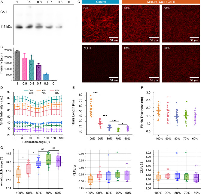
Collagen is critical to the structure and function of skin tissues, with the collagen I/III ratios influencing fibrillogenesis, fiber organization, and skin mechanics. Abnormal collagen organization, such as in fibrosis or scar tissue, compromises both skin functionality and aesthetics. In this study, we employed label-free polarization resolved second harmonic generation (PSHG) microscopy to investigate collagen structure in artificial collagen matrices with various Col I/III ratios at the fibril scale ( 1 to ) and in ex vivo human healthy and scarred skin at the fiber scale ( to ). Complementary third harmonic generation (THG) microscopy provided additional structural information. Our results indicate that an increasing Col I/III ratio is associated with longer fibril length, higher PSHG intensity, and a reduced effective -helix pitch angle of fibrils. In pure Col I, the effective -helix pitch angle is determined to be . These observations indicate alterations in fibril assembly. Furthermore, although the -helix pitch angle of fibers in both healthy and scarred skin was approximately , healthy skin exhibited greater variability in fiber orientation, suggesting a more randomized organization compared to scar tissue. THG imaging further revealed a higher cellular density in scar tissue, consistent with the inflammatory activity associated with wound healing. Immunohistochemical (IHC) staining using dermatansulphate and Col III-specific antibodies confirmed that the Col I/III ratio is higher in healthy skin (2.2) than in scarred skin (1.6). These findings underscore the potential of PSHG microscopy for label-free, quantitative assessment of collagen structure across multiple scales, with THG offering complementary cellular insights. This integrated approach represents a promising strategy for real-time, in vivo monitoring and automated quantification of collagen organization in clinical applications, including dermatology, burn treatment, and fibrosis monitoring.
胶原蛋白对于皮肤组织的结构和功能至关重要,I型/III型胶原蛋白的比例会影响原纤维形成、纤维组织和皮肤力学性能。胶原蛋白组织异常,如在纤维化或瘢痕组织中,会损害皮肤功能和美观。在本研究中,我们采用无标记偏振分辨二次谐波产生(PSHG)显微镜,在原纤维尺度(1至 )下研究具有不同I型/III型胶原蛋白比例的人工胶原蛋白基质中的胶原蛋白结构,并在纤维尺度( 至 )下研究离体人类健康皮肤和瘢痕皮肤中的胶原蛋白结构。互补的三次谐波产生(THG)显微镜提供了额外的结构信息。我们的结果表明,I型/III型胶原蛋白比例增加与原纤维长度增加、PSHG强度升高以及原纤维有效α-螺旋螺距角减小有关。在纯I型胶原蛋白中,有效α-螺旋螺距角确定为 。这些观察结果表明原纤维组装发生了改变。此外,尽管健康皮肤和瘢痕皮肤中纤维的α-螺旋螺距角约为 ,但健康皮肤在纤维取向方面表现出更大的变异性,这表明与瘢痕组织相比组织结构更随机。THG成像进一步揭示瘢痕组织中细胞密度更高,这与伤口愈合相关的炎症活动一致。使用硫酸皮肤素和III型胶原蛋白特异性抗体进行的免疫组织化学(IHC)染色证实,健康皮肤中的I型/III型胶原蛋白比例(2.2)高于瘢痕皮肤(1.6)。这些发现强调了PSHG显微镜在跨多个尺度对胶原蛋白结构进行无标记、定量评估方面的潜力,而THG提供了互补的细胞见解。这种综合方法代表了一种有前景的策略,可用于临床应用(包括皮肤病学、烧伤治疗和纤维化监测)中胶原蛋白组织的实时、体内监测和自动定量。