Rautou Pierre-Emmanuel, Chotkoe Shivani, Biquard Louise, Wettstein Guillaume, van der Graaff Denise, Liu Yao, De Man Joris, Casteleyn Christophe, Thys Sofie, De Vos Winnok H, Bedossa Pierre, Cooreman Michael P, Baudin Martine, Abitbol Jean-Louis, Huot-Marchand Philippe, Dzen Lucile, Albuquerque Miguel, Broqua Pierre, Junien Jean-Louis, Vonghia Luisa, Abdelmalek Manal F, Kwanten Wilhelmus J, Paradis Valérie, Francque Sven M
Université Paris-Cité, Inserm, Centre de recherche sur l'inflammation, Paris, France.
AP-HP, Hôpital Beaujon, Service d'Hépatologie, DMU DIGEST, Centre de Référence des Maladies Vasculaires du Foie, FILFOIE, ERN RARE-LIVER, Clichy, France.
JHEP Rep. 2025 Feb 22;7(6):101366. doi: 10.1016/j.jhepr.2025.101366. eCollection 2025 Jun.
BACKGROUND & AIMS: Data on changes in liver sinusoidal endothelial cells (LSECs) in patients with metabolic dysfunction-associated steatotic liver disease (MASLD) and their response to treatment are limited. This study aimed at determining (i) features associated with LSEC capillarisation in patients with MASLD; (ii) whether LSEC changes can regress with the pan-peroxisome proliferator-activated receptor (PPAR) agonist lanifibranor; (iii) the role of the different PPAR isotypes on LSEC changes in MASLD.
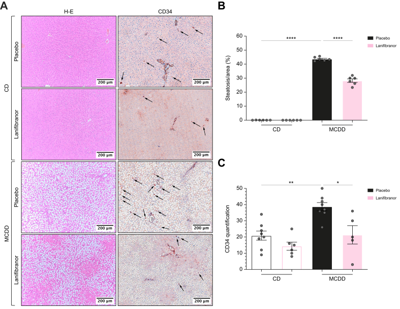
We analysed CD34 expression, a marker of LSEC capillarisation, on liver biopsies from patients considered for inclusion in the NATIVE trial at baseline (n = 249), and after 24 weeks of placebo or lanifibranor (n = 173). Two rat models of MASLD were used to investigate the effect of lanifibranor or of mono-PPAR agonists on LSECs.
Lobular CD34 staining was more intense in patients with isolated steatosis than in those with no MASLD (52% 10%; = 0.03). In the overall cohort, this staining was more intense in patients with metabolic dysfunction-associated steatohepatitis (MASH) than in those without (63% 41%; = 0.01) and strongly correlated with liver fibrosis and to a lesser extent with liver inflammation. Lanifibranor treatment was associated with more common improvement in CD34 periportal staining ( = 0.025), and less frequent worsening of lobular staining ( = 0.028). Compared with healthy rats, rats with MASLD had higher CD34 staining, portal venous pressure, intrahepatic vascular resistance, and impaired liver endothelial function. Lanifibranor normalised or strongly improved these abnormalities, whereas mono-PPAR agonists caused partial improvements.
In patients, LSEC capillarisation was increased at the earliest stages of MASLD and was associated with liver fibrosis and inflammation. In both patients and rats with MASLD, lanifibranor treatment was associated with improvement in liver endothelial phenotype.
Data on changes in liver sinusoidal endothelial cells (LSECs) in patients with metabolic dysfunction-associated steatotic liver disease (MASLD) and their response to treatment are limited. This study demonstrates that LSEC capillarisation is already present in the lobular zone of the liver of patients and rats at the stage of isolated steatosis, before metabolic dysfunction-associated steatohepatitis (MASH) onset, and progresses with liver fibrosis, and to a lesser extent with liver inflammation. Lanifibranor treatment, a pan-peroxisome proliferator-activated receptor agonist currently tested in a phase III clinical trial, improves LSEC capillarisation but also intrahepatic vascular resistance and portal pressure in MASLD. Targeting LSECs appears to be a promising approach to improve MASH.
关于代谢功能障碍相关脂肪性肝病(MASLD)患者肝窦内皮细胞(LSEC)变化及其对治疗反应的数据有限。本研究旨在确定:(i)MASLD患者中与LSEC毛细血管化相关的特征;(ii)LSEC变化是否可通过全过氧化物酶体增殖物激活受体(PPAR)激动剂拉尼贝特回归正常;(iii)不同PPAR亚型在MASLD的LSEC变化中的作用。
我们分析了在基线时(n = 249)考虑纳入NATIVE试验的患者肝活检组织中LSEC毛细血管化标志物CD34的表达,以及在接受安慰剂或拉尼贝特治疗24周后(n = 173)的表达情况。使用两种MASLD大鼠模型来研究拉尼贝特或单一PPAR激动剂对LSEC的影响。
孤立性脂肪变性患者的小叶CD34染色比无MASLD患者更强烈(52% ± 10%;P = 0.03)。在整个队列中,代谢功能障碍相关脂肪性肝炎(MASH)患者的这种染色比无MASH患者更强烈(63% ± 41%;P = 0.01),并且与肝纤维化密切相关,与肝脏炎症的相关性较小。拉尼贝特治疗与门静脉周围CD34染色更常见的改善相关(P = 0.025),小叶染色恶化的频率更低(P = 0.028)。与健康大鼠相比,MASLD大鼠的CD34染色、门静脉压力、肝内血管阻力更高,肝内皮功能受损。拉尼贝特使这些异常恢复正常或显著改善,而单一PPAR激动剂仅产生部分改善。
在患者中,LSEC毛细血管化在MASLD的最早阶段就增加了,并且与肝纤维化和炎症相关。在MASLD患者和大鼠中,拉尼贝特治疗均与肝内皮表型的改善相关。
关于代谢功能障碍相关脂肪性肝病(MASLD)患者肝窦内皮细胞(LSEC)变化及其对治疗反应的数据有限。本研究表明,在孤立性脂肪变性阶段,即在代谢功能障碍相关脂肪性肝炎(MASH)发作之前,患者和大鼠肝脏的小叶区域就已经存在LSEC毛细血管化,并且随着肝纤维化进展,与肝脏炎症的相关性较小。拉尼贝特治疗,一种目前正在III期临床试验中测试的全过氧化物酶体增殖物激活受体激动剂,可改善MASLD中的LSEC毛细血管化,还可改善肝内血管阻力和门静脉压力。靶向LSEC似乎是改善MASH的一种有前景的方法。