• 文献检索
  • 文档翻译
  • 深度研究
  • 学术资讯
  • Suppr Zotero 插件Zotero 插件
  • 邀请有礼
  • 套餐&价格
  • 历史记录
应用&插件
Suppr Zotero 插件Zotero 插件浏览器插件Mac 客户端Windows 客户端微信小程序
定价
高级版会员购买积分包购买API积分包
服务
文献检索文档翻译深度研究API 文档MCP 服务
关于我们
关于 Suppr公司介绍联系我们用户协议隐私条款
关注我们

Suppr 超能文献

核心技术专利:CN118964589B侵权必究
粤ICP备2023148730 号-1Suppr @ 2026

文献检索

告别复杂PubMed语法,用中文像聊天一样搜索,搜遍4000万医学文献。AI智能推荐,让科研检索更轻松。

立即免费搜索

文件翻译

保留排版,准确专业,支持PDF/Word/PPT等文件格式,支持 12+语言互译。

免费翻译文档

深度研究

AI帮你快速写综述,25分钟生成高质量综述,智能提取关键信息,辅助科研写作。

立即免费体验

泡型包虫病的超声诊断:最新进展

Ultrasound Diagnostics in Alveolar Echinococcosis: An Update.

作者信息

Kratzer Wolfgang

机构信息

Department of Internal Medicine I, University Hospital Ulm, Ulm, Germany.

出版信息

Visc Med. 2025 May 3:1-7. doi: 10.1159/000545080.

DOI:10.1159/000545080
PMID:40486189
原文链接:https://pmc.ncbi.nlm.nih.gov/articles/PMC12143886/
Abstract

BACKGROUND

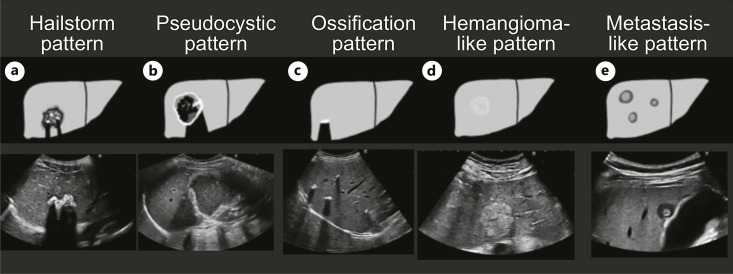
Alveolar echinococcosis (AE) is a rare, potentially fatal zoonosis. In recent years, imaging diagnostics have become increasingly important compared to serologic diagnostics in AE. We will provide an overview of the importance of ultrasound diagnostics in AE in the detection of the disease and its significance in follow-up, as well as the typical sonographic presentation patterns and pitfalls.

SUMMARY

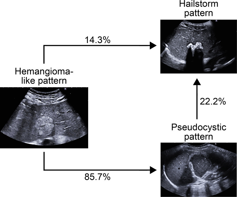
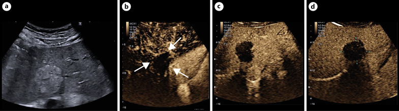
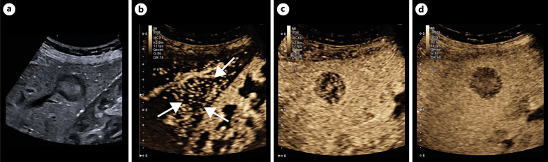
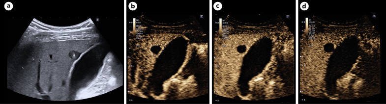
The use of the ultrasound classification developed by us and the use of contrast-enhanced ultrasound (CEUS) helps make the diagnosis faster and better. Without CEUS, the hemangioma-like pattern and the metastasis-like pattern in particular cannot be diagnosed with certainty. The limitations of ultrasound diagnostics on the patient side, in terms of examiner experience and equipment, remain.

KEY MESSAGES

Ultrasound is an important procedure in the detection and follow-up of AE. Contrast-enhanced sonography is indispensable. Fundamental limitations of ultrasound diagnostics such as examiner experience remain.

摘要

背景

肺泡型棘球蚴病(AE)是一种罕见的、潜在致命的人畜共患病。近年来,与AE的血清学诊断相比,影像学诊断变得越来越重要。我们将概述超声诊断在AE疾病检测中的重要性及其在随访中的意义,以及典型的超声表现模式和陷阱。

总结

使用我们开发的超声分类方法以及对比增强超声(CEUS)有助于更快、更好地进行诊断。没有CEUS,特别是血管瘤样模式和转移样模式无法确诊。在患者方面,超声诊断在检查者经验和设备方面的局限性仍然存在。

关键信息

超声是AE检测和随访中的重要检查方法。对比增强超声检查不可或缺。超声诊断的基本局限性,如检查者经验,仍然存在。

https://cdn.ncbi.nlm.nih.gov/pmc/blobs/9357/12143886/c3bd4c0a2dae/vis-2025-0000-0000-545080_F07.jpg
https://cdn.ncbi.nlm.nih.gov/pmc/blobs/9357/12143886/1224eb8c5a2b/vis-2025-0000-0000-545080_F01.jpg
https://cdn.ncbi.nlm.nih.gov/pmc/blobs/9357/12143886/22de0549d0c0/vis-2025-0000-0000-545080_F02.jpg
https://cdn.ncbi.nlm.nih.gov/pmc/blobs/9357/12143886/99f4055d78e1/vis-2025-0000-0000-545080_F03.jpg
https://cdn.ncbi.nlm.nih.gov/pmc/blobs/9357/12143886/726084b94f49/vis-2025-0000-0000-545080_F04.jpg
https://cdn.ncbi.nlm.nih.gov/pmc/blobs/9357/12143886/e066a3cf7009/vis-2025-0000-0000-545080_F05.jpg
https://cdn.ncbi.nlm.nih.gov/pmc/blobs/9357/12143886/0bbc3944e2d4/vis-2025-0000-0000-545080_F06.jpg
https://cdn.ncbi.nlm.nih.gov/pmc/blobs/9357/12143886/c3bd4c0a2dae/vis-2025-0000-0000-545080_F07.jpg

相似文献

1
Ultrasound Diagnostics in Alveolar Echinococcosis: An Update.泡型包虫病的超声诊断:最新进展
Visc Med. 2025 May 3:1-7. doi: 10.1159/000545080.
2
Long-term follow-up of liver alveolar echinococcosis using echinococcosis multilocularis ultrasound classification.肝泡型包虫病的超声分类及其长期随访。
World J Gastroenterol. 2021 Oct 28;27(40):6939-6950. doi: 10.3748/wjg.v27.i40.6939.
3
Differentiation of hepatic alveolar echinococcosis with a hemangioma-like pattern compared to typical liver hemangioma using contrast-enhanced ultrasound: a pilot study.应用超声造影对比分析肝泡型包虫病与典型肝血管瘤的鉴别诊断:一项初步研究。
Infection. 2023 Feb;51(1):159-168. doi: 10.1007/s15010-022-01866-z. Epub 2022 Jul 1.
4
The use of contrast-enhanced ultrasound (CEUS) for the diagnostic evaluation of hepatic echinococcosis.超声造影(CEUS)在肝包虫病诊断评估中的应用。
Clin Hemorheol Microcirc. 2018;70(4):449-455. doi: 10.3233/CH-189310.
5
Contrast-enhanced ultrasound (CEUS) in patients with metastasis-like hepatic alveolar echinococcosis: a cohort study.伴有转移样表现的肝泡型包虫病患者的超声造影:一项队列研究。
J Ultrasound. 2023 Mar;26(1):129-136. doi: 10.1007/s40477-022-00688-x. Epub 2022 May 21.
6
Can contrast-enhanced ultrasound differentiate the type of hepatic echinococcosis: cystic echinococcosis or alveolar echinococcosis?增强超声能否区分肝包虫病的类型:肝囊型包虫病或泡型包虫病?
Parasit Vectors. 2023 Apr 17;16(1):131. doi: 10.1186/s13071-023-05731-2.
7
Ultrasonographic findings of small lesion of hepatic alveolar echinococcosis.肝泡型包虫病小病灶的超声检查结果
Acta Trop. 2017 Oct;174:165-170. doi: 10.1016/j.actatropica.2016.01.030. Epub 2016 Feb 15.
8
Comparison of ultrasonographic features between two types of hepatic echinococcosis and hepatoblastoma in children.儿童两种肝包虫病与肝母细胞瘤超声特征的比较。
Front Pediatr. 2025 Jan 28;13:1458649. doi: 10.3389/fped.2025.1458649. eCollection 2025.
9
Recent advances in ultrasound in the diagnosis and evaluation of the activity of hepatic alveolar echinococcosis.超声在肝泡型包虫病诊断和活动评估中的最新进展。
Parasitol Res. 2021 Sep;120(9):3077-3082. doi: 10.1007/s00436-021-07262-0. Epub 2021 Aug 9.
10
Ultrasound 2020 - Diagnostics & Therapy: On the Way to Multimodal Ultrasound: Contrast-Enhanced Ultrasound (CEUS), Microvascular Doppler Techniques, Fusion Imaging, Sonoelastography, Interventional Sonography.超声 2020 - 诊断与治疗:迈向多模态超声之路:超声造影(CEUS)、微血管多普勒技术、融合成像、声弹性成像、介入性超声。
Rofo. 2021 Jan;193(1):23-32. doi: 10.1055/a-1217-7400. Epub 2020 Jul 30.

本文引用的文献

1
Major complications after ultrasound-guided liver biopsy: An annual audit of a Chinese tertiary-care teaching hospital.超声引导下肝活检术后的主要并发症:一家中国三级甲等教学医院的年度审计
World J Gastrointest Surg. 2023 Jul 27;15(7):1388-1396. doi: 10.4240/wjgs.v15.i7.1388.
2
Can contrast-enhanced ultrasound differentiate the type of hepatic echinococcosis: cystic echinococcosis or alveolar echinococcosis?增强超声能否区分肝包虫病的类型:肝囊型包虫病或泡型包虫病?
Parasit Vectors. 2023 Apr 17;16(1):131. doi: 10.1186/s13071-023-05731-2.
3
[Diagnostic delaying factors in hepatic alveolar echinococcosis].
[肝泡型包虫病的诊断延误因素]
Dtsch Med Wochenschr. 2023 Apr;148(8):e37-e43. doi: 10.1055/a-1996-3603. Epub 2023 Jan 23.
4
Dog Ownership and Risk for Alveolar Echinococcosis, Germany.养狗与泡型包虫病风险,德国。
Emerg Infect Dis. 2022 Aug;28(8):1597-1605. doi: 10.3201/eid2808.212514.
5
Differentiation of hepatic alveolar echinococcosis with a hemangioma-like pattern compared to typical liver hemangioma using contrast-enhanced ultrasound: a pilot study.应用超声造影对比分析肝泡型包虫病与典型肝血管瘤的鉴别诊断:一项初步研究。
Infection. 2023 Feb;51(1):159-168. doi: 10.1007/s15010-022-01866-z. Epub 2022 Jul 1.
6
Contrast-enhanced ultrasound (CEUS) in patients with metastasis-like hepatic alveolar echinococcosis: a cohort study.伴有转移样表现的肝泡型包虫病患者的超声造影:一项队列研究。
J Ultrasound. 2023 Mar;26(1):129-136. doi: 10.1007/s40477-022-00688-x. Epub 2022 May 21.
7
Role of the radiologist in the diagnosis and management of the two forms of hepatic echinococcosis.放射科医生在两种肝包虫病的诊断与管理中的作用。
Insights Imaging. 2022 Apr 8;13(1):68. doi: 10.1186/s13244-022-01190-y.
8
18F-FDG-PET/MR in Alveolar Echinococcosis: Multiparametric Imaging in a Real-World Setting.18F-FDG-PET/MR在肺泡型棘球蚴病中的应用:真实世界中的多参数成像
Pathogens. 2022 Mar 12;11(3):348. doi: 10.3390/pathogens11030348.
9
Echinococcosis: a Challenge for Liver Sonography.包虫病:肝脏超声检查的挑战。
Ultraschall Med. 2022 Apr;43(2):120-145. doi: 10.1055/a-1694-5552. Epub 2022 Feb 24.
10
Long-term follow-up of liver alveolar echinococcosis using echinococcosis multilocularis ultrasound classification.肝泡型包虫病的超声分类及其长期随访。
World J Gastroenterol. 2021 Oct 28;27(40):6939-6950. doi: 10.3748/wjg.v27.i40.6939.