Hoveizi Elham, Naddaf Hadi, Naeimavi Emad
Department of Biology, Faculty of Science, Shahid Chamran University of Ahvaz, Ahvaz, Iran.
Department of Clinical Sciences, Faculty of Veterinary, Shahid Chamran University of Ahvaz, Ahvaz, Iran.
Regen Ther. 2025 May 15;30:31-45. doi: 10.1016/j.reth.2025.05.003. eCollection 2025 Dec.
This research aims to use neural-like cells (NLCs) derived from endometrial mesenchymal stem cells (EnMSCs) on a polylactic acid/chitosan scaffold (PLA/CS) along with the use of (Memocopa) in a rat sciatic nerve injury model for sciatic nerve regeneration. While previous studies have explored stem cell therapies and scaffold-based approaches for nerve regeneration, using EnMSCs in combination with a PLA/CS scaffold and Memocopa represents a novel, potentially synergistic approach.
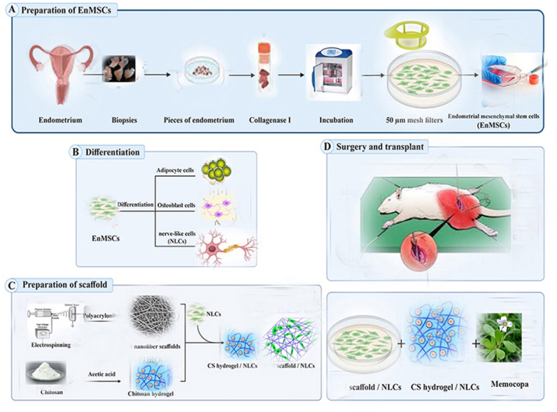
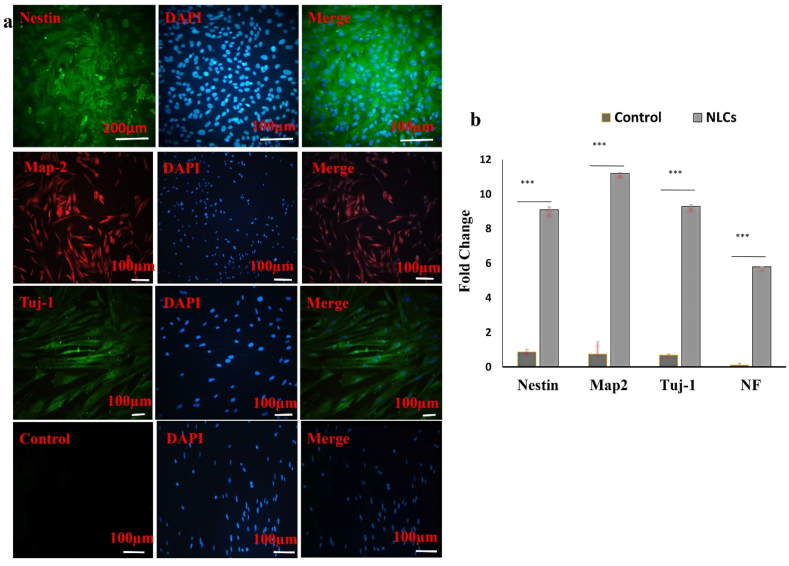
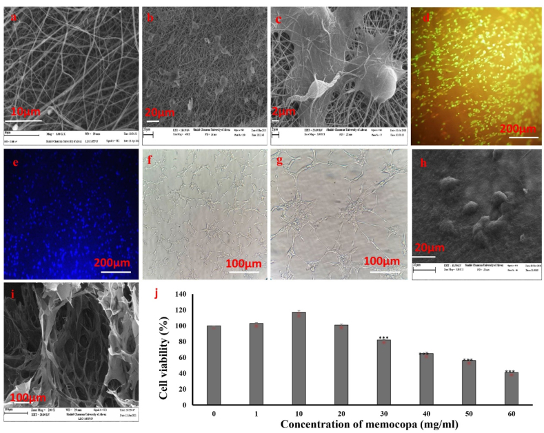
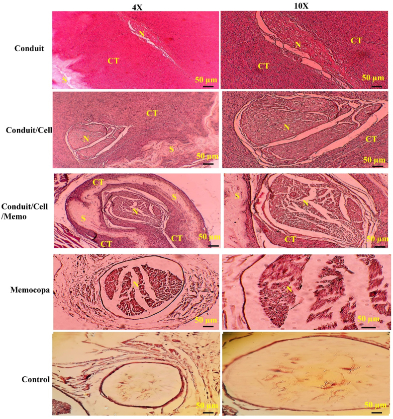
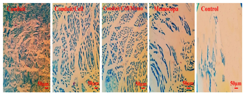
EnMSCs were isolated, characterized, and differentiated in a study. The expression of specific genes in the differentiated cells was confirmed using RT-PCR and immunocytochemistry. PLA nanofiber and chitosan hydrogel scaffolds were created for neural tissue engineering. Memocopa was administered orally alongside scaffold and cell transplantation. The study involved 25 adult male Wistar rats with a 3 mm sciatic nerve gap, divided into five groups based on treatment. Animals were monitored for 8 weeks, during which SFI was measured. Tissue samples were then prepared for histological examination, including various staining techniques.
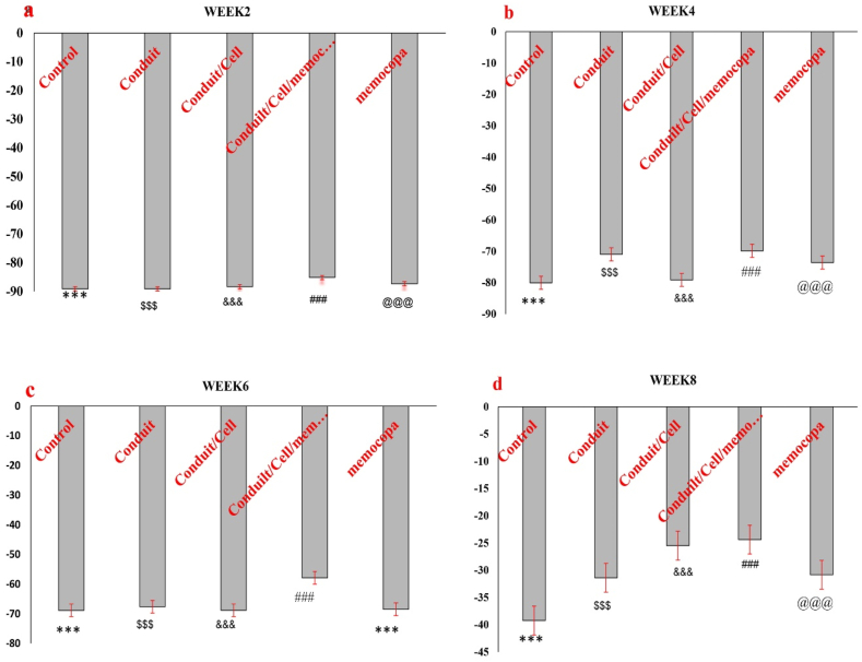
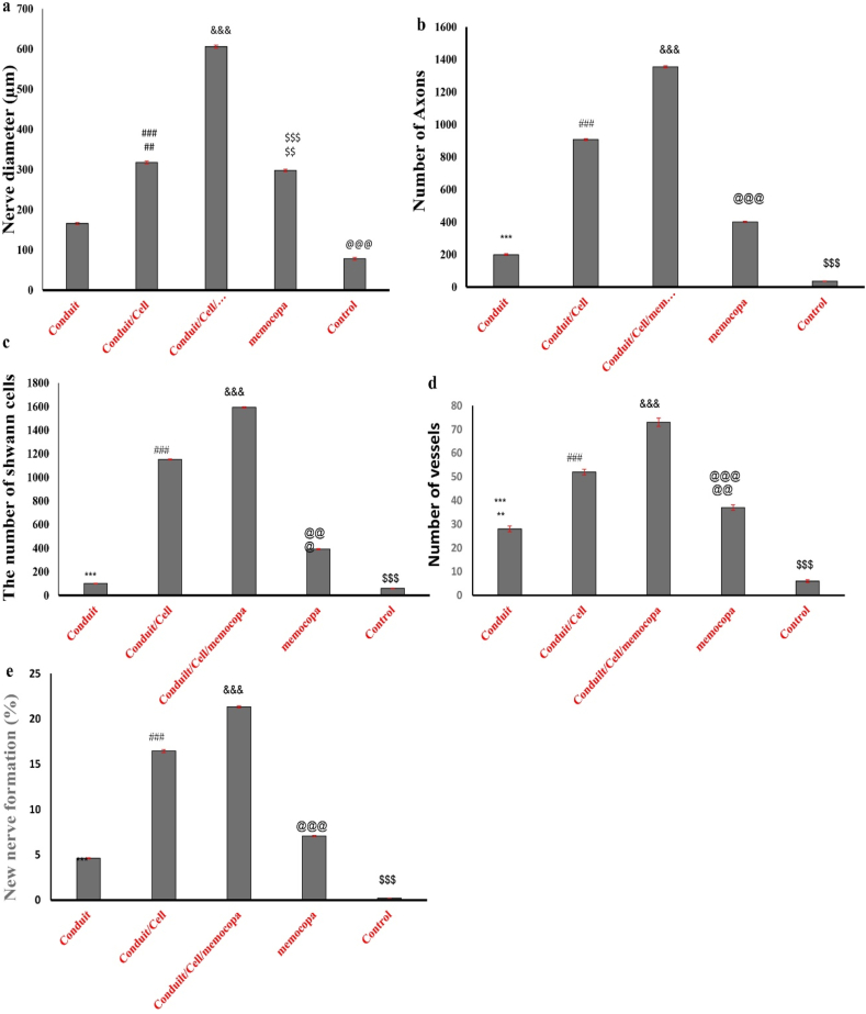
The combination of scaffold, cells, and Memocopa showed significant improvements in sciatic nerve function, as indicated by the SFI results in the eighth week: conduit group -33.87, conduit/cells group -25.92, conduit/cells/Memocopa group -22.86, Memocopa group -30.93, and control group -38.87. Histological findings revealed improvements in various aspects, including percentages of new nerve formation across the different treatment groups: conduit group 4.62 %, conduit/cells group 16.45 %, conduit/cells/Memocopa group 21.32 %, Memocopa group 7.07 %, and control group 0.22 %.
The results of this study showed that efficient differentiation of EnMSCs into NLCs is possible, and with the help of PLA/CS scaffold and simultaneous use of Memocopa, it is possible to repair and improve sciatic nerve injury in a rat animal model.
本研究旨在将源自子宫内膜间充质干细胞(EnMSCs)的神经样细胞(NLCs)应用于聚乳酸/壳聚糖支架(PLA/CS),并在大鼠坐骨神经损伤模型中联合使用(Memocopa)促进坐骨神经再生。虽然先前的研究已经探索了干细胞疗法和基于支架的神经再生方法,但将EnMSCs与PLA/CS支架和Memocopa联合使用代表了一种新颖的、可能具有协同作用的方法。
在一项研究中分离、鉴定并诱导EnMSCs分化。使用逆转录聚合酶链反应(RT-PCR)和免疫细胞化学法确认分化细胞中特定基因的表达。制备用于神经组织工程的PLA纳米纤维和壳聚糖水凝胶支架。在支架和细胞移植的同时口服Memocopa。该研究涉及25只成年雄性Wistar大鼠,其坐骨神经有3毫米的间隙,根据治疗方法分为五组。对动物进行8周的监测,在此期间测量坐骨神经功能指数(SFI)。然后制备组织样本用于组织学检查,包括各种染色技术。
支架、细胞和Memocopa的联合使用在坐骨神经功能方面显示出显著改善,如第八周的SFI结果所示:导管组-33.87,导管/细胞组-25.92,导管/细胞/Memocopa组-22.86,Memocopa组-30.93,对照组-38.87。组织学结果显示在各个方面都有改善,包括不同治疗组新神经形成的百分比:导管组4.62%,导管/细胞组16.45%,导管/细胞/Memocopa组21.32%,Memocopa组7.07%,对照组0.22%。
本研究结果表明,EnMSCs高效分化为NLCs是可能的,并且在PLA/CS支架的帮助下同时使用Memocopa,可以修复和改善大鼠动物模型中的坐骨神经损伤。