Jiang Wang, Yin Jie, Han Min, He Wenhua, Zhao Yan, Hu Jinyue, Wang Meiwei, Wang Sikai, Xu Jiafan, Deng Chongtian, Li Jiajia, Gong Xiaonuo, Shen Yueming
Department of Digestive Diseases, The Affiliated Changsha Central Hospital, Hengyang Medical School, University of South China, Changsha, 410000, People's Republic of China.
Department of Cardiovascular Diseases, The Affiliated Changsha Central Hospital, Hengyang Medical School, University of South China, Changsha, 410000, People's Republic of China.
J Inflamm Res. 2025 Jun 3;18:7167-7181. doi: 10.2147/JIR.S518155. eCollection 2025.
N4BP3 is a ubiquitination-related gene that plays a pivotal role in neurology and neoplasia. Studies have demonstrated its essential function in axonal and dendritic branching, promoting hepatocellular carcinoma and breast cancer. Our previous research reveals that N4BP3 enhances inflammatory responses by modulating the NOD2 signaling pathway. It is crucial to investigate whether N4BP3 regulates inflammatory bowel disease (IBD) through the TLR4 signaling pathway and to elucidate the underlying mechanisms.
Lipopolysaccharides (LPS) were used to activate the TLR4 pathway in THP-1/Caco-2 cells. THP-1/Caco-2 cells were transfected with either N4BP3 overexpression or knockdown plasmids, generating N4BP3-overexpressing or N4BP3-deficient cell lines. For in vivo studies, colitis was induced in mice using dextran sodium sulfate (DSS). Additionally, negative control and N4BP3-knockdown C57BL/6 mouse models were established via intraperitoneal injection of control or N4BP3-targeting adeno-associated virus (AAV).
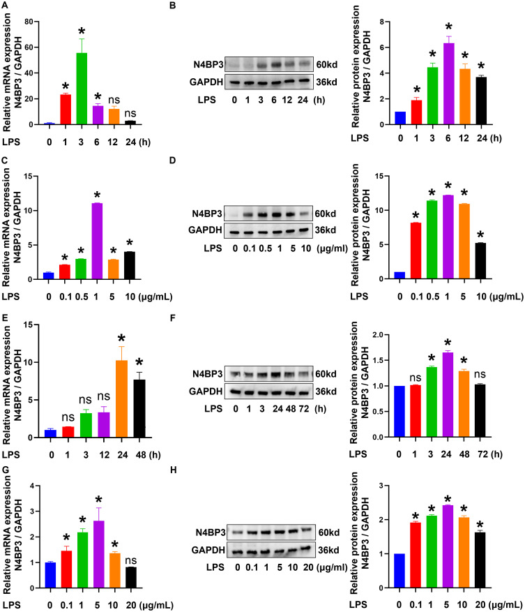
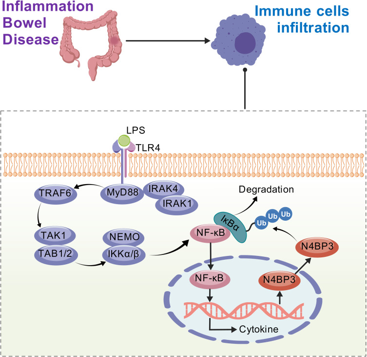
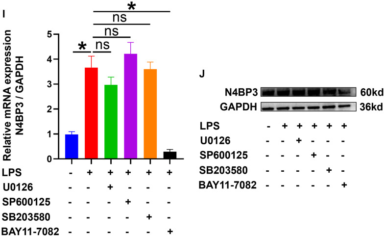
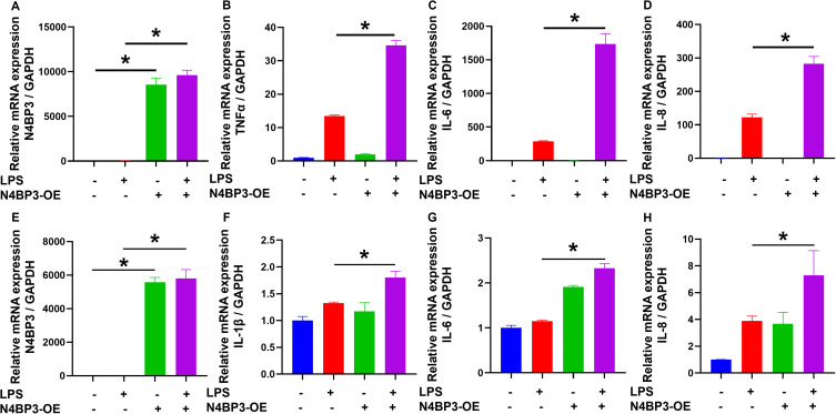
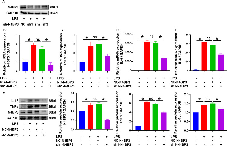
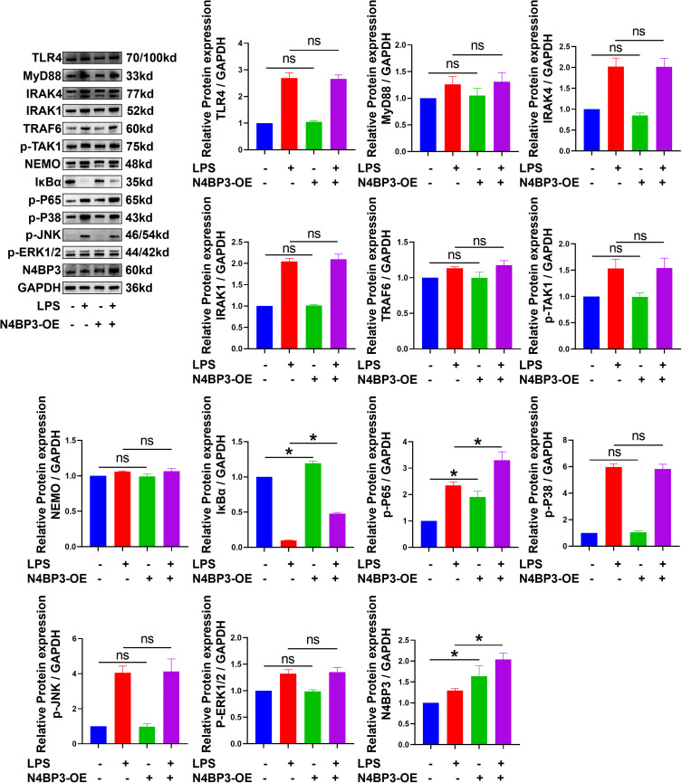
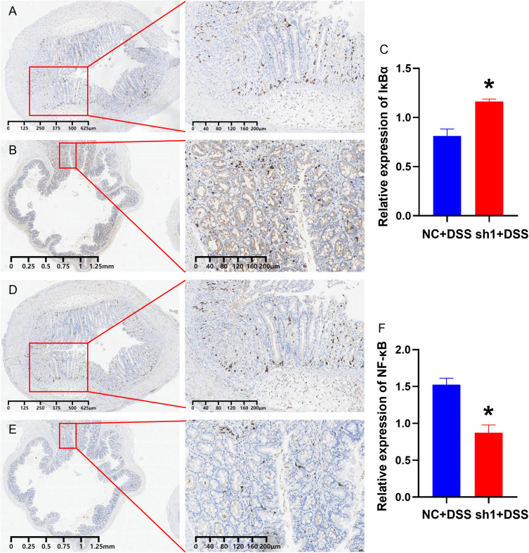
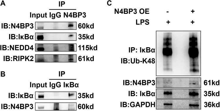
LPS stimulation significantly upregulated N4BP3 expression in THP-1/Caco-2 cells compared to sterile water treatment (P < 0.05). In N4BP3-overexpressing cells, LPS induction led to significantly higher expression of TNF-α, IL-1β, IL-6, and IL-8 mRNA, as well as phospho-NF-κB p65 protein, compared to wild-type THP-1/Caco-2 cells (P < 0.05). Conversely, these inflammatory markers were markedly downregulated in N4BP3-knockdown THP-1 cells following LPS stimulation (P < 0.05). In DSS-induced colitis models, N4BP3-knockdown mice showed decreased phospho-NF-κB p65 but increased IκBα protein expression in colonic tissues compared to DSS-treated control mice (P < 0.05). Furthermore, we observed interaction between N4BP3 and IκBα, with N4BP3-overexpressing THP-1 cells demonstrating significantly elevated K48-linked ubiquitination levels versus controls.
LPS upregulates N4BP3 expression, which subsequently enhances K48-linked ubiquitination of IκBα, leading to NF-κB pathway activation, and exacerbating IBD progression. These findings suggest N4BP3 as a potential therapeutic target for developing novel IBD treatments.
N4BP3是一个与泛素化相关的基因,在神经学和肿瘤形成中起关键作用。研究已证明其在轴突和树突分支、促进肝细胞癌和乳腺癌方面的重要功能。我们之前的研究表明,N4BP3通过调节NOD2信号通路增强炎症反应。研究N4BP3是否通过Toll样受体4(TLR4)信号通路调节炎症性肠病(IBD)并阐明其潜在机制至关重要。
使用脂多糖(LPS)激活THP-1/Caco-2细胞中的TLR4通路。用N4BP3过表达或敲低质粒转染THP-1/Caco-2细胞,构建N4BP3过表达或N4BP3缺陷细胞系。在体内研究中,使用葡聚糖硫酸钠(DSS)诱导小鼠发生结肠炎。此外,通过腹腔注射对照或靶向N4BP3的腺相关病毒(AAV)建立阴性对照和N4BP3敲低的C57BL/6小鼠模型。
与无菌水处理相比,LPS刺激显著上调了THP-1/Caco-2细胞中N4BP3的表达(P<0.05)。与野生型THP-1/Caco-2细胞相比,在N4BP3过表达细胞中,LPS诱导导致肿瘤坏死因子-α(TNF-α)、白细胞介素-1β(IL-1β)、白细胞介素-6(IL-6)和白细胞介素-8(IL-8)mRNA以及磷酸化核因子-κB p65蛋白的表达显著更高(P<0.05)。相反,在LPS刺激后,N4BP3敲低的THP-1细胞中这些炎症标志物明显下调(P<0.05)。在DSS诱导的结肠炎模型中,与DSS处理的对照小鼠相比,N4BP3敲低的小鼠结肠组织中磷酸化核因子-κB p65减少,但IκBα蛋白表达增加(P<0.05)。此外,我们观察到N4BP3与IκBα之间存在相互作用,与对照相比,N4BP3过表达的THP-1细胞中K48连接的泛素化水平显著升高。
LPS上调N4BP3表达,随后增强IκBα的K48连接的泛素化,导致核因子-κB通路激活,并加剧IBD进展。这些发现表明N4BP3是开发新型IBD治疗方法的潜在治疗靶点。