Zainul Abidin Fatin N, Biondo Francesca, Altmann Andre, Dawson Sally J
UCL Ear Institute, University College London, London WC1X 8EE, UK.
UCL Hawkes Institute, Department of Medical Physics and Biomedical Engineering, University College London, London WC1V 6BH, UK.
Brain Commun. 2025 May 27;7(3):fcaf203. doi: 10.1093/braincomms/fcaf203. eCollection 2025.
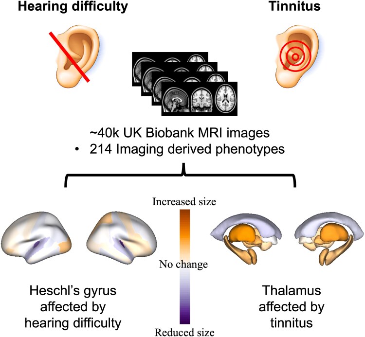
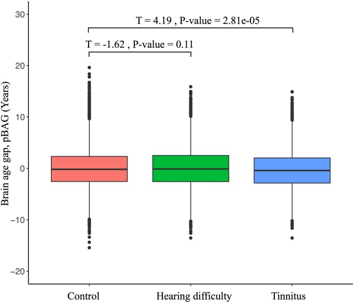
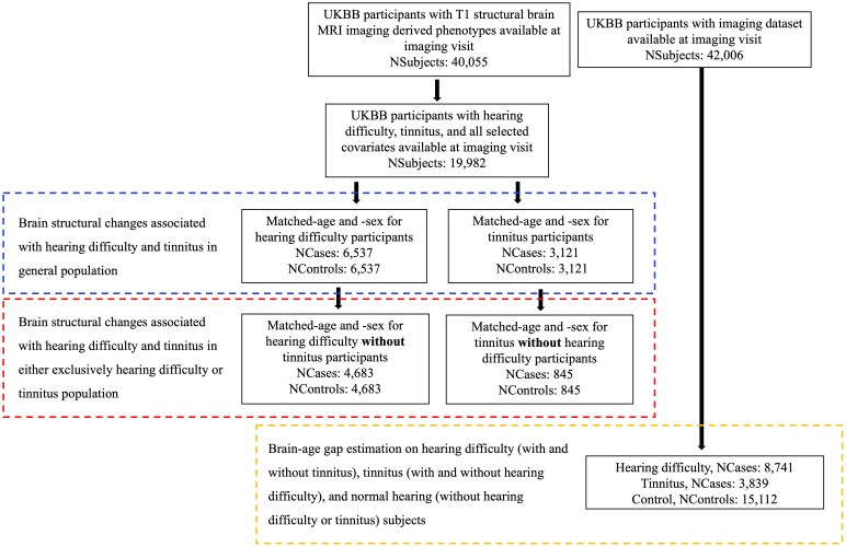
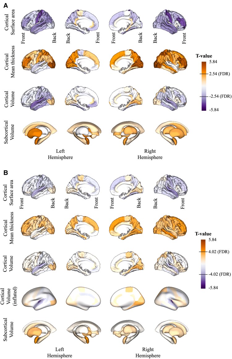
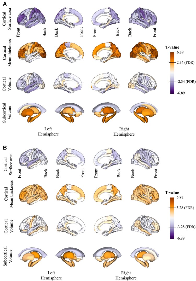
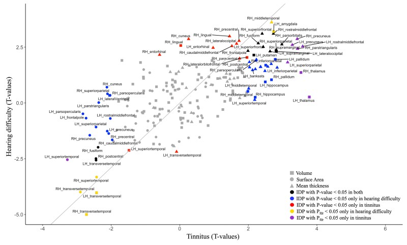
Prevalence of both hearing loss and tinnitus increases with age. However, neuroimaging studies of both conditions report inconsistent changes in brain morphology likely due to small sample size and variable methodology. Structural and functional neuroimaging studies in hearing loss and tinnitus have revealed distinct neural correlates, and further replication is needed to confirm these findings. This study aims to investigate the effects of hearing loss and tinnitus on the brain morphology in a well-powered sample. We utilized self-reported hearing difficulty and tinnitus in participants with magnetic resonance imaging (MRI) in the UK Biobank cohort. Control participants without hearing difficulty and tinnitus were age and sex matched leading to total sample sizes of 13 074 and 6242 for self-reported hearing difficulty and tinnitus, respectively. We utilized the rich UK Biobank dataset (i) to reveal these brain changes in a well-powered large study of hearing loss and tinnitus, (ii) to document the effect of confounding factors on these associations, (iii) to discriminate the effects of tinnitus versus hearing difficulty on the brain and (iv) to estimate the brain-age gap in hearing difficulty and tinnitus subjects compared with controls. Hearing difficulty is significantly associated with smaller grey matter volumes exclusively in the bilateral transverse temporal regions, whereas tinnitus is associated with larger volumes of bilateral hippocampi and thalami when compared with the control group. Furthermore, correcting for confounders (i.e. diabetes, cardiovascular disease, age, sex, smoking, alcohol consumption and Townsend deprivation index) during statistical analysis helped to better delineate the impact of hearing status on brain structural changes. The brain-age gap analysis showed that participants with tinnitus appeared to have significantly younger brains than controls, whereas participants with hearing difficulty did not differ significantly from the control group. Altogether, our results confirmed previous findings and suggest the enlargement of bilateral thalami as the main effect in people with tinnitus. We also established that there are independent and distinct brain pathologies between hearing difficulty and tinnitus. Therefore, the self-reported measure is a reasonable approach to assess the hearing loss and tinnitus pathologies.
听力损失和耳鸣的患病率均随年龄增长而增加。然而,针对这两种情况的神经影像学研究报告称,由于样本量小和方法多变,大脑形态的变化并不一致。听力损失和耳鸣的结构及功能神经影像学研究已揭示出不同的神经关联,需要进一步重复研究以证实这些发现。本研究旨在通过一个样本量充足的样本,调查听力损失和耳鸣对大脑形态的影响。我们在英国生物银行队列中,利用参与者自我报告的听力困难和耳鸣情况,并结合磁共振成像(MRI)进行研究。无听力困难和耳鸣的对照参与者在年龄和性别上进行了匹配,自我报告听力困难和耳鸣的总样本量分别为13074例和6242例。我们利用丰富的英国生物银行数据集:(i)在一项样本量充足的听力损失和耳鸣大型研究中揭示这些大脑变化;(ii)记录混杂因素对这些关联的影响;(iii)区分耳鸣与听力困难对大脑的影响;(iv)估计听力困难和耳鸣受试者与对照组相比的脑龄差距。听力困难仅与双侧颞横回区域灰质体积减小显著相关,而与对照组相比,耳鸣与双侧海马体和丘脑体积增大相关。此外,在统计分析过程中校正混杂因素(即糖尿病、心血管疾病、年龄、性别、吸烟、饮酒和汤森贫困指数)有助于更好地描绘听力状况对脑结构变化的影响。脑龄差距分析表明,耳鸣参与者的大脑似乎比对照组明显更年轻,而听力困难参与者与对照组无显著差异。总之,我们的结果证实了先前的发现,并表明双侧丘脑增大是耳鸣患者的主要影响因素。我们还确定,听力困难和耳鸣之间存在独立且不同的脑部病变。因此,自我报告测量是评估听力损失和耳鸣病变的一种合理方法。