• 文献检索
  • 文档翻译
  • 深度研究
  • 学术资讯
  • Suppr Zotero 插件Zotero 插件
  • 邀请有礼
  • 套餐&价格
  • 历史记录
应用&插件
Suppr Zotero 插件Zotero 插件浏览器插件Mac 客户端Windows 客户端微信小程序
定价
高级版会员购买积分包购买API积分包
服务
文献检索文档翻译深度研究API 文档MCP 服务
关于我们
关于 Suppr公司介绍联系我们用户协议隐私条款
关注我们

Suppr 超能文献

核心技术专利:CN118964589B侵权必究
粤ICP备2023148730 号-1Suppr @ 2026

文献检索

告别复杂PubMed语法,用中文像聊天一样搜索,搜遍4000万医学文献。AI智能推荐,让科研检索更轻松。

立即免费搜索

文件翻译

保留排版,准确专业,支持PDF/Word/PPT等文件格式,支持 12+语言互译。

免费翻译文档

深度研究

AI帮你快速写综述,25分钟生成高质量综述,智能提取关键信息,辅助科研写作。

立即免费体验

石蜡包埋组织与块面成像的精确MRI-组织学配准

Precise MRI-Histology Coregistration of Paraffin-Embedded Tissue with Blockface Imaging.

作者信息

Wang Yixin, Ho William, Huszar Istvan N, DiGiacomo Phillip, Taghavi Hossein Moein, Tao Lee, Choi Matthew, Nguyen Nhu, Leventis Samantha, Camarillo David B, Schlömer Philipp, Axer Markus, Wei Shao, Rusu Mirabela, Cobos Inma, Nirschl Jeff, Georgiadis Marios, Zeineh Michael

机构信息

Department of Bioengineering, Stanford University, Stanford, CA, USA.

Department of Radiology, Stanford School of Medicine, Stanford, CA, USA.

出版信息

bioRxiv. 2025 Jun 5:2025.06.02.657335. doi: 10.1101/2025.06.02.657335.

DOI:10.1101/2025.06.02.657335
PMID:40501733
原文链接:https://pmc.ncbi.nlm.nih.gov/articles/PMC12157374/
Abstract

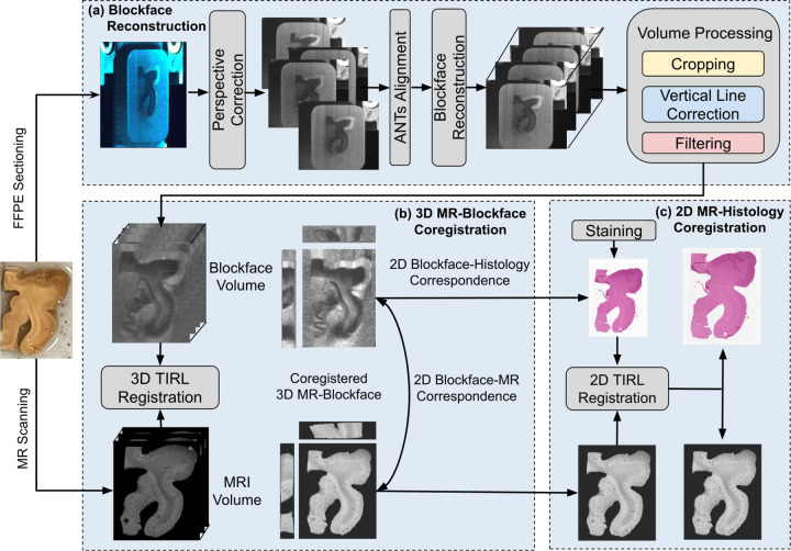
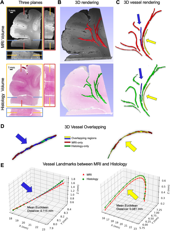
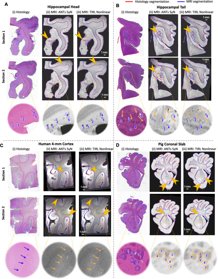
Magnetic resonance imaging (MRI) provides 3D spatial information on tissue, yet it lacks at the molecular level. In contrast, histology provides cellular and molecular information, but it lacks the 3D spatial context and direct translation. Coregistering the two is key for the 3D-embedding of histological details, validating pathological MRI findings, and finding quantitative imaging biomarkers of neurodegenerative diseases. However, coregistration is challenging due to non-linear distortions of the tissue from histological processing and sectioning leading to microscopic and macroscopic nonlinear 3D deformations between specimen MRI and stained histology sections. To address this, we developed a novel pipeline, named Brewster's Blockface Quantification (BBQ), integrating robust optical approaches with innovative 2D and 3D registration algorithms to achieve precise volumetric alignment of specimen MRI data with histological images. On a variety of brain tissue specimens from distinct anatomical regions and across multiple species, our methodology generated blockface volumes with minimal distortion and artifacts. Using these blockface volumes as an intermediary, we achieve a precise alignment between MRI and histology slides, yielding registration results with an overlapping Dice score of ~90% for whole tissue alignment between MRI and blockface volumes, and >95% for 2D MRI-histology registration. This correlative MRI-histology pipeline with robust 2D and 3D coregistration methods promises to enhance our understanding of neurodegenerative diseases and aid the development of MRI-based disease biomarkers.

摘要

磁共振成像(MRI)可提供组织的三维空间信息,但在分子层面存在不足。相比之下,组织学能提供细胞和分子信息,却缺乏三维空间背景及直接的转化。将两者进行配准是对组织学细节进行三维嵌入、验证MRI病理结果以及寻找神经退行性疾病定量成像生物标志物的关键。然而,由于组织学处理和切片过程中组织的非线性变形,导致标本MRI与染色组织学切片之间出现微观和宏观的非线性三维变形,配准具有挑战性。为解决这一问题,我们开发了一种名为布鲁斯特块面量化(BBQ)的新型流程,将强大的光学方法与创新的二维和三维配准算法相结合,以实现标本MRI数据与组织学图像的精确体积对齐。在来自不同解剖区域和多个物种的各种脑组织标本上,我们的方法生成的块面体积失真和伪影最小。以这些块面体积作为中介,我们实现了MRI与组织学切片之间的精确对齐,在MRI与块面体积之间的全组织对齐中,重叠Dice分数约为90%,在二维MRI - 组织学配准中大于95%。这种具有强大二维和三维配准方法的相关MRI - 组织学流程有望加深我们对神经退行性疾病的理解,并有助于基于MRI的疾病生物标志物的开发。

https://cdn.ncbi.nlm.nih.gov/pmc/blobs/e08f/12157374/3ca4b266c9f2/nihpp-2025.06.02.657335v1-f0012.jpg
https://cdn.ncbi.nlm.nih.gov/pmc/blobs/e08f/12157374/0bb1673ab2d3/nihpp-2025.06.02.657335v1-f0001.jpg
https://cdn.ncbi.nlm.nih.gov/pmc/blobs/e08f/12157374/8ecd0700f558/nihpp-2025.06.02.657335v1-f0002.jpg
https://cdn.ncbi.nlm.nih.gov/pmc/blobs/e08f/12157374/8fa3a11d4646/nihpp-2025.06.02.657335v1-f0003.jpg
https://cdn.ncbi.nlm.nih.gov/pmc/blobs/e08f/12157374/862a2b9fa692/nihpp-2025.06.02.657335v1-f0004.jpg
https://cdn.ncbi.nlm.nih.gov/pmc/blobs/e08f/12157374/9210646348ca/nihpp-2025.06.02.657335v1-f0005.jpg
https://cdn.ncbi.nlm.nih.gov/pmc/blobs/e08f/12157374/dd602e94e6a0/nihpp-2025.06.02.657335v1-f0006.jpg
https://cdn.ncbi.nlm.nih.gov/pmc/blobs/e08f/12157374/ef557e360abb/nihpp-2025.06.02.657335v1-f0007.jpg
https://cdn.ncbi.nlm.nih.gov/pmc/blobs/e08f/12157374/ae2b82722545/nihpp-2025.06.02.657335v1-f0008.jpg
https://cdn.ncbi.nlm.nih.gov/pmc/blobs/e08f/12157374/ca3546bf8173/nihpp-2025.06.02.657335v1-f0009.jpg
https://cdn.ncbi.nlm.nih.gov/pmc/blobs/e08f/12157374/741ead58465a/nihpp-2025.06.02.657335v1-f0010.jpg
https://cdn.ncbi.nlm.nih.gov/pmc/blobs/e08f/12157374/d6a7562559af/nihpp-2025.06.02.657335v1-f0011.jpg
https://cdn.ncbi.nlm.nih.gov/pmc/blobs/e08f/12157374/3ca4b266c9f2/nihpp-2025.06.02.657335v1-f0012.jpg

相似文献

1
Precise MRI-Histology Coregistration of Paraffin-Embedded Tissue with Blockface Imaging.石蜡包埋组织与块面成像的精确MRI-组织学配准
bioRxiv. 2025 Jun 5:2025.06.02.657335. doi: 10.1101/2025.06.02.657335.
2
Precise MRI-histology coregistration of paraffin-embedded tissue with blockface imaging.石蜡包埋组织与块面成像的精确MRI-组织学配准。
Imaging Neurosci (Camb). 2025 Aug 8;3. doi: 10.1162/IMAG.a.106. eCollection 2025.
3
Prescription of Controlled Substances: Benefits and Risks管制药品的处方:益处与风险
4
An open-source deep learning framework for respiratory motion monitoring and volumetric imaging during radiation therapy.一种用于放射治疗期间呼吸运动监测和容积成像的开源深度学习框架。
Med Phys. 2025 Jul;52(7):e18015. doi: 10.1002/mp.18015.
5
Magnetic resonance perfusion for differentiating low-grade from high-grade gliomas at first presentation.首次就诊时磁共振灌注成像用于鉴别低级别与高级别胶质瘤
Cochrane Database Syst Rev. 2018 Jan 22;1(1):CD011551. doi: 10.1002/14651858.CD011551.pub2.
6
Influence of early through late fusion on pancreas segmentation from imperfectly registered multimodal magnetic resonance imaging.早期至晚期融合对来自配准不完善的多模态磁共振成像的胰腺分割的影响。
J Med Imaging (Bellingham). 2025 Mar;12(2):024008. doi: 10.1117/1.JMI.12.2.024008. Epub 2025 Apr 26.
7
Intraoperative frozen section analysis for the diagnosis of early stage ovarian cancer in suspicious pelvic masses.术中冰冻切片分析用于诊断可疑盆腔肿块中的早期卵巢癌。
Cochrane Database Syst Rev. 2016 Mar 1;3(3):CD010360. doi: 10.1002/14651858.CD010360.pub2.
8
Development of histological sectioning techniques for the cochlear implanted inner ear in miniature swine.小型猪人工耳蜗植入内耳组织切片技术的发展
J Neurosci Methods. 2025 Jul 7;423:110527. doi: 10.1016/j.jneumeth.2025.110527.
9
Bridging Neuroimaging and Neuropathology: A Comprehensive Workflow for Targeted Sampling of White Matter Lesions.连接神经影像学与神经病理学:白质病变靶向采样的综合工作流程
medRxiv. 2025 Jun 9:2025.06.08.25329217. doi: 10.1101/2025.06.08.25329217.
10
Aspects of Genetic Diversity, Host Specificity and Public Health Significance of Single-Celled Intestinal Parasites Commonly Observed in Humans and Mostly Referred to as 'Non-Pathogenic'.人类常见且大多被称为“非致病性”的单细胞肠道寄生虫的遗传多样性、宿主特异性及公共卫生意义
APMIS. 2025 Sep;133(9):e70036. doi: 10.1111/apm.70036.