Farhat Nadim, Li Jinghang, Berardinelli Jacob, Stauffer Mark, Sajewski Andrea, Alkhateeb Salem, Schweitzer Noah, Hecheng Jin, Ikonomovic Milos, Liou Jr-Jiun, Aizenstein Howard J, Mettenburg Joseph, Santini Tales, Wu Minjie, Kofler Julia, Ibrahim Tamer S
University of Pittsburgh, Department of Bioengineering, Pittsburgh, PA, US.
University of Pittsburgh School of Medicine, Department of Pathology, Pittsburgh, PA, US.
medRxiv. 2025 Jun 9:2025.06.08.25329217. doi: 10.1101/2025.06.08.25329217.
White matter lesions are common imaging biomarkers associated with aging and neurodegenerative diseases, yet their underlying pathology remains unclear due to limitations in imaging-based characterization. We aim to develop and validate a comprehensive workflow enabling precise MRI-guided histological sampling of white matter lesions to bridge neuroimaging and neuropathology.
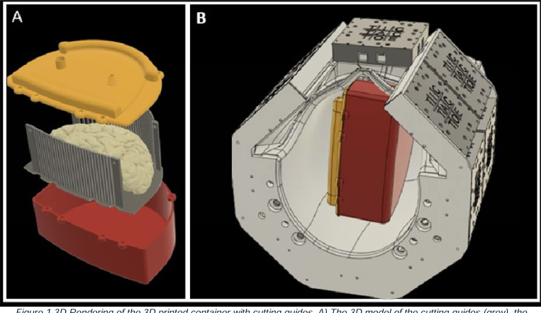
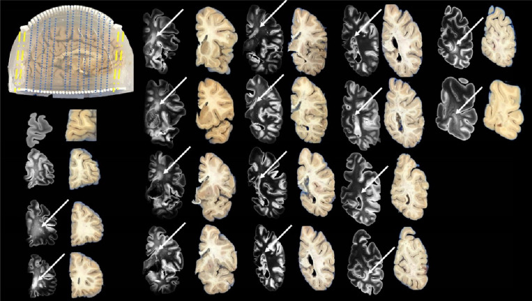
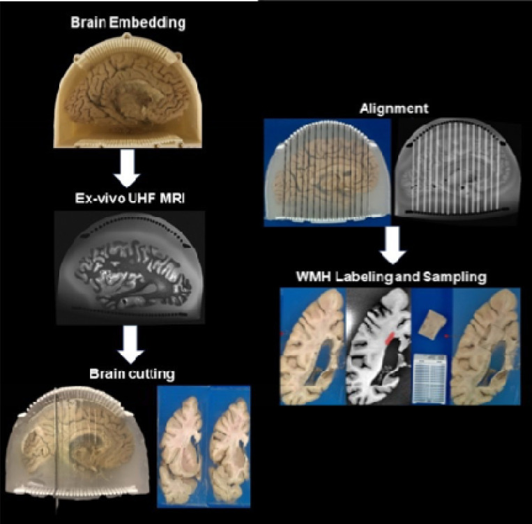
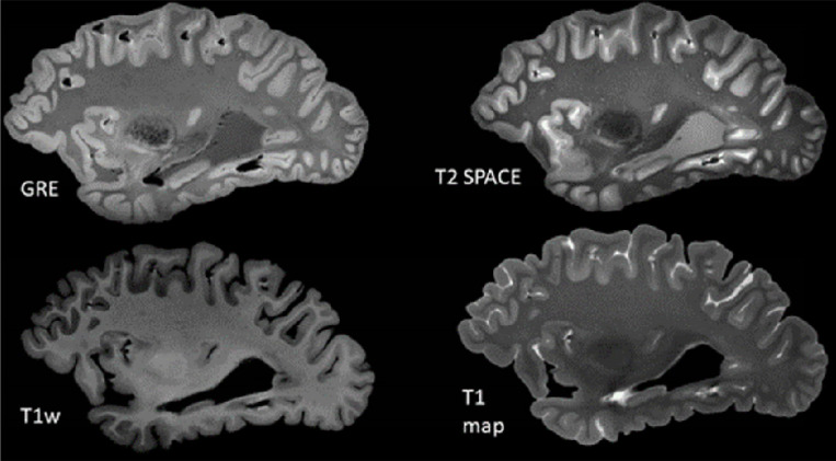
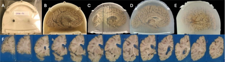
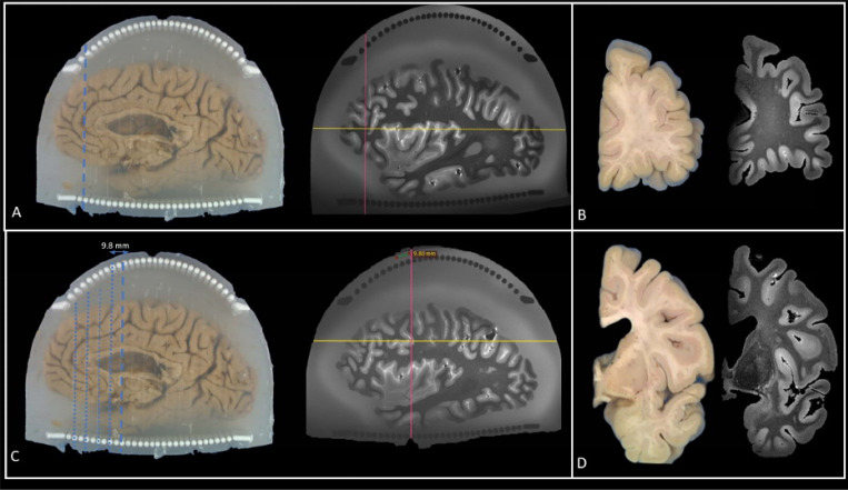
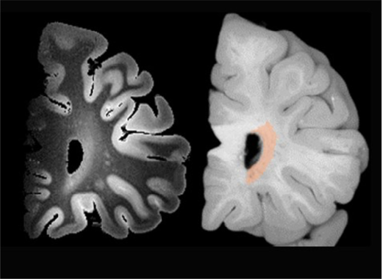
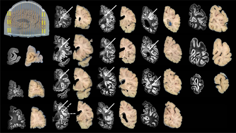
We establish a workflow integrating agarose-saccharose brain embedding, ultra-high field 7T MRI acquisition, reusable 3D-printed cutting guides, and semi-automated MRI-blockface alignment. Postmortem brains are stabilized in the embedding medium and scanned using optimized MRI protocols. Coronal sectioning is guided by standardized 3D-printed cutting guides, and knife traces are digitally matched to MRI planes. White matter lesions are segmented on MRI and aligned for histopathological sampling. This approach is validated in over 100 postmortem human brains.
The workflow enables reproducible brain sectioning, minimizes imaging artifacts, and achieves precise spatial alignment between MRI and histology. Consistent, high-resolution MRI data facilitated accurate lesion detection and sampling. The use of standardized cutting guides and alignment protocols reduce variability and improve efficiency.
Our cost-effective, scalable workflow reliably links neuroimaging findings with histological analysis, enhancing the understanding of white matter lesion pathology. This framework holds significant potential for advancing translational research in aging and neurodegenerative diseases.
白质病变是与衰老和神经退行性疾病相关的常见影像学生物标志物,但由于基于成像的特征描述存在局限性,其潜在病理学仍不清楚。我们旨在开发并验证一种全面的工作流程,实现对白质病变进行精确的MRI引导下组织学采样,以弥合神经影像学和神经病理学之间的差距。
我们建立了一个工作流程,整合了琼脂糖-蔗糖脑包埋、超高场7T MRI采集、可重复使用的3D打印切割导向器和半自动MRI-脑块面配准。死后大脑在包埋介质中固定,并使用优化的MRI协议进行扫描。冠状切片由标准化的3D打印切割导向器引导,刀痕与MRI平面进行数字匹配。在MRI上对白质病变进行分割,并进行配准以进行组织病理学采样。该方法在100多例死后人类大脑中得到验证。
该工作流程能够实现可重复的脑切片,最大限度地减少成像伪影,并在MRI和组织学之间实现精确的空间配准。一致的高分辨率MRI数据有助于准确检测和采样病变。使用标准化的切割导向器和配准协议可减少变异性并提高效率。
我们具有成本效益、可扩展的工作流程可靠地将神经影像学发现与组织学分析联系起来,增强了对白质病变病理学的理解。该框架在推进衰老和神经退行性疾病的转化研究方面具有巨大潜力。