Boor P J, Ferrans V J
Am J Pathol. 1985 Oct;121(1):39-54.
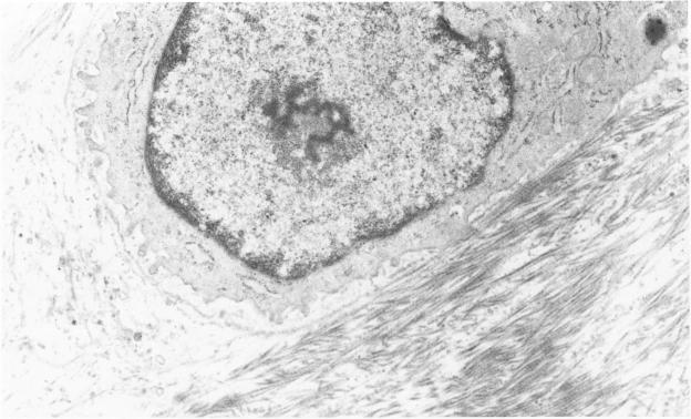
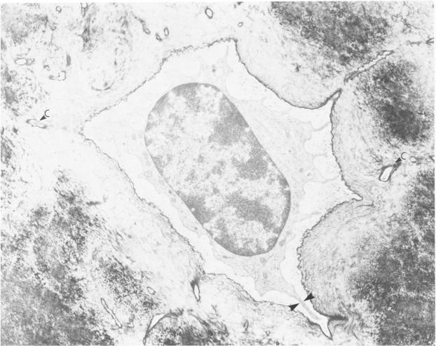
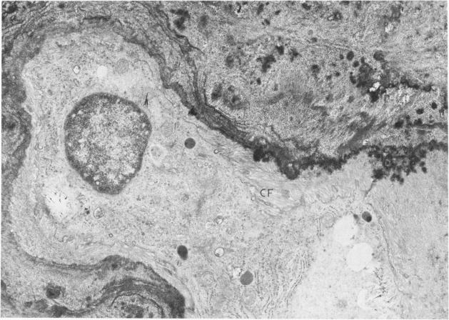
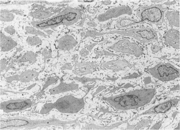
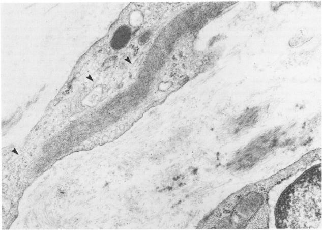
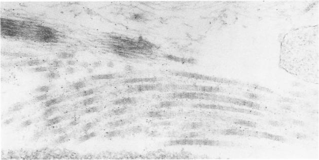
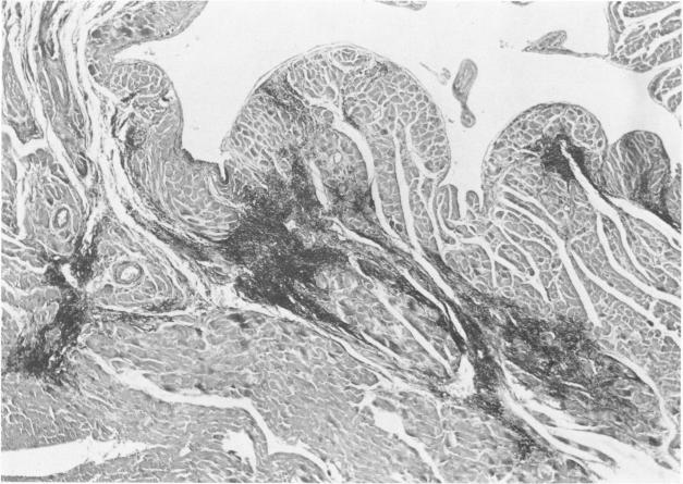
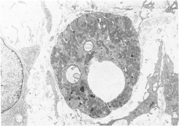
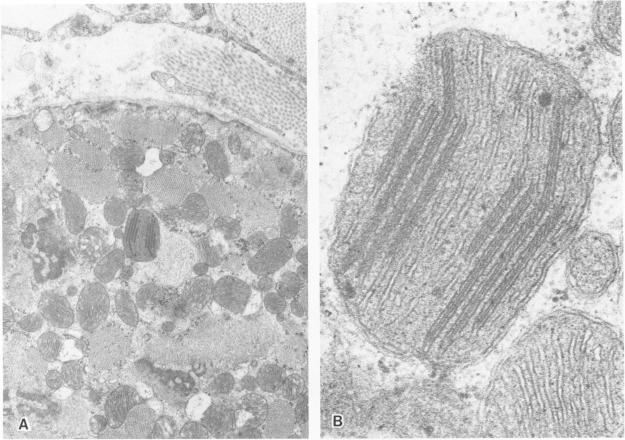
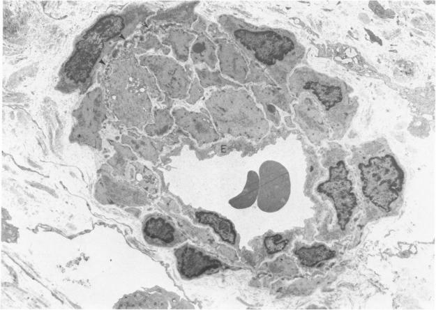
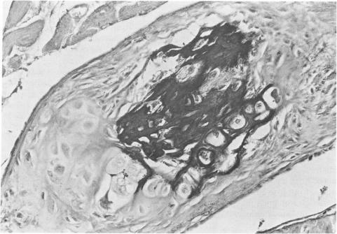
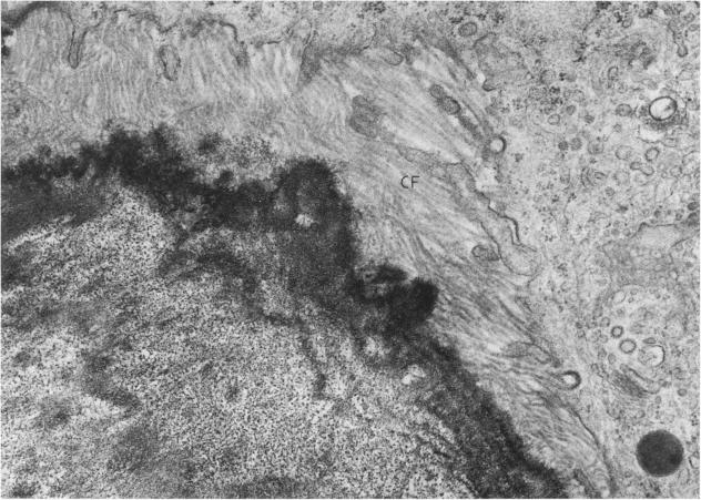
The late myocardial and vascular ultrastructural changes in rat hearts following consumption of the cardiovascular toxin allylamine were studied. Rats were given 0.1% allylamine HCl in drinking water for 10-104 days. From 10 to 21 days, there was organization of acute myocardial necrosis by macrophages and scattered polymorphonuclear leukocytes with prominent interstitial-cell proliferation. Alterations at 21-104 days included extensive scarring with formation of dense mature collagen with scattered fibroblasts present, grossly evident left-ventricular aneurysm, and gross and microscopic changes similar to those observed in the secondary form of endocardial fibroelastosis. Areas of scar contained highly cellular foci of smooth-muscle cells, myofibroblasts, and abundant extracellular elastin. Cardiac myocytes frequently showed markedly disorganized myofilaments, bizarrely distorted mitochondria with condensed cristae, and other severe degenerative changes. Small vessels within and adjacent to scar showed proliferation of intimal smooth-muscle cells. Endothelial lesions or recent or organized thrombi were not seen. Focal endocardial metaplasia, consisting of both chondroid and osseous tissue, was found in areas of transmural scarring, or ventricular aneurysm. Chondrocytes had the overall nuclear and cellular morphology, abundant rough endoplasmic reticulum, and surrounding lacunae typical of mature fibrocartilage. In some areas, the collagen matrix was undergoing calcification with the typical cross-banded pattern of calcifying connective tissue. Osteocytes were located in a densely calcified bone matrix and displayed characteristic cellular extensions into surrounding canaliculi. These findings indicate a severe myocardial, small-vessel, and endocardial injury during the course of chronic allylamine intoxication.
研究了大鼠摄入心血管毒素烯丙胺后心脏晚期的心肌和血管超微结构变化。给大鼠饮用含0.1%烯丙胺盐酸盐的水10 - 104天。在10至21天,急性心肌坏死由巨噬细胞和成簇的多形核白细胞进行机化,伴有明显的间质细胞增殖。在21至104天的改变包括广泛的瘢痕形成,有致密成熟胶原形成,存在散在的成纤维细胞,肉眼可见左心室动脉瘤,以及大体和显微镜下的变化类似于心内膜弹力纤维增生症的继发性形式。瘢痕区域含有平滑肌细胞、肌成纤维细胞高度聚集的灶以及丰富的细胞外弹性蛋白。心肌细胞常表现出肌丝明显紊乱、线粒体异常扭曲且嵴浓缩,以及其他严重的退行性变化。瘢痕内及瘢痕附近的小血管显示内膜平滑肌细胞增殖。未见内皮病变或近期形成或机化的血栓。在透壁性瘢痕或心室动脉瘤区域发现了由软骨样组织和骨组织组成的局灶性心内膜化生。软骨细胞具有成熟纤维软骨典型的核和细胞形态、丰富的粗面内质网以及周围的陷窝。在一些区域,胶原基质正在钙化,具有钙化结缔组织典型的交叉带状模式。骨细胞位于致密钙化的骨基质中,并显示出特征性的细胞突起延伸至周围的小管中。这些发现表明在慢性烯丙胺中毒过程中存在严重的心肌、小血管和心内膜损伤。