Keeratitanont Keattichai, Limchareon Sornsupha
Radiology and Nuclear Medicine, Burapha University Faculty of Medicine, Chonburi, Thailand
Radiology and Nuclear Medicine, Burapha University Faculty of Medicine, Chonburi, Thailand.
BMJ Open. 2025 Jun 13;15(6):e087424. doi: 10.1136/bmjopen-2024-087424.
To examine the association between body mass index (BMI) categories and the fragility fractures in Thai men and to identify the most common anatomical sites of these fractures. We hypothesised that BMI is associated with the risk of fragility fractures in this population.
Retrospective observational study.
A tertiary care centre in eastern Thailand, based on data from Burapha University Hospital.
The study included 419 Thai men aged 40 years or older who underwent bone mineral density (BMD) assessment between 2014 and 2022. Participants were classified according to the presence or absence of documented fragility fractures. Exclusion criteria included pathological fractures, high-energy trauma and incomplete BMI or BMD data.
The primary outcome was the association between BMI categories and the risk of fragility fractures. The secondary outcome was the anatomical distribution of these fractures.
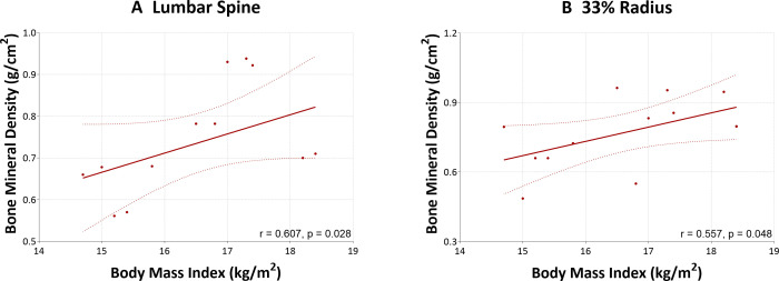
Among 419 participants, 147 (35.1%) had fragility fractures and 272 (64.9%) did not. Underweight men had significantly increased odds of fragility fractures (OR, 3.44; 95% CI, 1.03 to 11.47; p=0.044) and vertebral fractures (OR, 4.30; 95% CI, 1.36 to 13.58; p=0.013), compared with men of normal BMI. In contrast, overweight men had lower odds of overall fractures (OR, 0.50; 95% CI, 0.31 to 0.80; p=0.004) and vertebral fractures (OR, 0.48; 95% CI, 0.27 to 0.84; p=0.010). Among underweight participants, BMI was moderately positively correlated with BMD at the lumbar spine (r=0.607; p=0.028) and at the one-third radius (r=0.557; p=0.084).
Lower BMI was significantly associated with increased risk of fragility fractures, particularly vertebral fractures. These findings support prior evidence in Asian populations and reveal a fracture pattern, predominantly vertebral followed by hip fractures, which differs from those observed in predominantly Caucasian populations.
研究泰国男性体重指数(BMI)类别与脆性骨折之间的关联,并确定这些骨折最常见的解剖部位。我们假设BMI与该人群脆性骨折的风险相关。
回顾性观察研究。
基于泰国东部一所三级医疗中心——武里南大学医院的数据。
该研究纳入了419名年龄在40岁及以上的泰国男性,他们在2014年至2022年间接受了骨密度(BMD)评估。参与者根据是否有记录的脆性骨折进行分类。排除标准包括病理性骨折、高能量创伤以及BMI或BMD数据不完整。
主要结果是BMI类别与脆性骨折风险之间的关联。次要结果是这些骨折的解剖分布。
在419名参与者中,147人(35.1%)发生了脆性骨折,272人(64.9%)未发生。与BMI正常的男性相比,体重过轻的男性发生脆性骨折的几率显著增加(比值比[OR],3.44;95%置信区间[CI],1.03至11.47;p = 0.044)以及椎体骨折的几率(OR,4.30;95% CI,1.36至13.58;p = 0.013)。相比之下,超重男性发生总体骨折的几率较低(OR,0.50;95% CI,0.3至0.80;p = 0.004)以及椎体骨折的几率(OR,0.48;95% CI,0.27至0.84;p = 0.010)。在体重过轻的参与者中,BMI与腰椎骨密度(r = 0.607;p = 0.028)和桡骨远端三分之一处骨密度(r = 0.557;p = 0.084)呈中度正相关。
较低的BMI与脆性骨折风险增加显著相关,尤其是椎体骨折。这些发现支持了亚洲人群先前的证据,并揭示了一种骨折模式,主要是椎体骨折,其次是髋部骨折,这与主要在白种人群中观察到的情况不同。